Archive for the ‘plant viruses’ Category

Maize streak virus: the early history

30 March, 2015

The history of maize streak virus research is generally taken as starting in 1901, with the publication of the

The cover of the "Fuller Report"

The cover of the “Fuller Report”

by “Claude Fuller, Entomologist”. However, in the Report he does make reference to articles in the “Agricultural Journal” for August 3rd and 31st, 1900, and quotes personal sources as having noticed the disease of “mealie variegation” as early as the 1870s.  He comments that:

“…mealie growers…have been acquainted with variegated mealies…for at least 20 years…”, and “…Thomas Kirkman…has known the disorder for 30 years past…”.

His conclusions, although carefully arrived at, were very wrong. Fuller claimed the disease was due to soil deficiency or a “chemical enzyme” in soils, and could be combatted by intensive cultivation and “chemical manures”. However, his carefully-written account is still of great historical interest, and the observations are valuable as they are objective accounts of a skilled scientist.  The records of streaked grasses in particular are useful, as we still collect such samples to this day.  Fuller was later sadly a victim of one the first traffic accidents in what was then Lourenco Marques in Mozambique.

Streak symptoms in a maize leaf

The disease – now known as maize streak disease (MSD) – occurs only in Africa and adjacent Indian Ocean islands, where it is one of the worst occurring in maize.  The causal agent was discovered to be a virus by HH Storey in 1932, who termed it maize streak virus (MSV). The virus was found to be obligately transmitted by the leafhopper Cicadulina mbila, also by Storey, in 1928. In 1978, MSV was designated the type virus of the newly described group taxon Geminivirus.

Early studies indicated that there were several distinctly different African streak viruses adapted to different host ranges (Storey & McClean, 1930; McClean, 1947). These studies were based on the transmission of virus isolates between different host species and symptomatology.

In a subsequent study of streak virus transmission between maize, sugarcane, and Panicum maximum, the relatively new technique of immunodiffusion was employed, using antiserum to the maize isolate. From the results it was concluded that the maize, sugarcane, and Panicum isolates were strains of the same virus, MSV (Bock et al., 1974). The maize isolate was given as the type strain. The virus was only properly physically characterised in 1974, when the characteristic geminate or doubled particles were first seen by electron microscopy, and only found to be a single-stranded circular DNA virus in 1977 (Harrison et al., 1977).

Maize streak virus: photo from Robert G Milne in 1978

Maize streak virus: photo from Robert G Milne in 1978

The first isolates of MSV were sequenced in 1984 (Kenya, S Howell, 1984; Nigeria, P Mullineaux et al., 1984), and the virus was found to have a single component of single-stranded circular DNA (sscDNA), and to be about 2700 bases in size. The two isolates were about 98% identical in sequence. The second team took delight in noting that the first sequence was in fact of the complementary and not the virion strand.

A major advance in the field occurred in 1987, when Nigel Grimsley et al. showed that a tandem dimer clone of MSV-N in an Agrobacterium tumefaciens Ti plasmid-derived cloning vector, was infectious when the bacterium was injected into maize seedlings. Subsequently, Sondra Lazarowitz (1988) obtained the sequence of an infectious clone of a South African isolate (from Potchefstroom) – MSV-SA – and showed that it also shared about 98% identity with the first two sequences.

Since the early days other transmission tests and more sophisticated serological assays were performed on a wide range of streak isolates from different hosts and locales, and it was claimed that all forms of streak disease in the Gramineae in Africa were caused by strains of the same virus, MSV. This view changed as more and more viruses were characterised, however, and it became obvious that there were distinctly separate groupings of viruses that constituted different species: these were sugarcane streak viruses (SSV, see Hughes et al., 1993), the panicum streak viruses (PanSV, see Briddon et al., 1992), and the maize streak viruses. Together these viruses constituted an African streak virus group (see Hughes et al., 1992; Rybicki and Hughes, 1990), distinct from an Australasian striate mosaic virus group, and other more distantly related viruses (see here for the state of the art in 1997).  These studies together with a later one by Rybicki et al. in 1998 also pointed up the utility of the polymerase chain reaction (PCR) for amplification, detection and subsequent sequencing of DNA from diverse mastreviruses.

A more modern and comprehensive account can also be found here, in a recent review written for Molecular Plant Pathology.

Back to Contents

Virology Africa 2015: consider yourselves notified!

7 November, 2014

Dear ViroBlogy and Virology News followers:

Anna-Lise Williamson and I plan to have another in our irregular series of “Virology Africa” conferences in November-December 2015, in Cape Town.

As previously, the conference will run over 3 days or so, possibly with associated workshops, and while the venue is not decided, we would like to base it at least partially in the Victoria & Alfred Waterfront.

We also intend to cover the whole spectrum of virology, from human through animal to plant; clinical aspects and biotechnology.

We intend to make it as cheap as possible so that students can come. We will also not be inviting a slate of international speakers, as we have found that we always get quite an impressive slate without having to fund them fully.

It is also the intention to have a Plant Molecular Farming workshop – concentrating on plant-made vaccines – concurrently with the conference, in order to leverage existing bilateral travel grants with international partners. If anyone else has such grants that could be similarly leveraged, it would be greatly appreciated.

See you in Cape Town in 2015!

Ed + Anna-Lise

How can geminiviral Rep capture the cell cycle of differentiated plant cells?

12 August, 2014

African cassava mosaic virus (ACMV) in the geminivirus family has being affected 500 million people worldwide by devastating cassava crops during the past decades. It has caused severe symptoms and reduced yield up to the complete loss of roots, the main starchy food source especially for subsistence farmers in Africa. How can a tiny virus with a small genome evoke such dramatic effects? The viral key component, the replication-initiator protein (Rep), forces differentiated plant cells in the phloem to reactivate DNA synthesis. Even more, it does the same in model cells of fission yeast. We have identified, now, a potential cyclin interaction motif, RXL, in the sequence of ACMV Rep, which may be important for cell cycle control. This motif is essential to induce rereplication in yeast and necessary for viral infection of plants.


Source: www.virologyhighlights.com

I am a sucker for geminiviruses and their replication – as can be seen in the pages published here and elsewhere over the years.  It is fascinating to me that a small protein like Rep – only ~30 kDa – can do so many things, and especially interfere in such a fundamental way with organised, differentiated cells.

What is even more interesting is that it can do it in such a wide variety of systems: it’s been shown that ACMV can replicate in maize protoplasts as well as in the dicotyledonous cassava; it can evidently function well in yeast as well – and via a pathway that no-one suspected before now.

Truly, a protein of many parts!  Congratulations to Katharina Hipp and to my old friends Bruno and Holger.

See on Scoop.itVirology News

A new virus from the Namib – and a guilty secret revealed

26 June, 2014

I have to confess to a guilty secret: there is a pleasure-inducing activity I have been indulging in for a week at a time these past three years.

And not alone….

This consists of going to the Gobabeb Research & Training Centre in the Namib Desert as part of an international “scientific expedition” aimed at investigating microbial soil biodiversity in the sandy and stony desert round Gobabeb.  These were started by Professor Don Cowan when he was at the University of the Western Cape, and have fortunately continued now that he has moved to a new Institute at the University of Pretoria.

Typical quartz-associated hypolith

Typical quartz-associated hypolith

I put the scientific expedition in quotes because anything that much fun shouldn’t be called scientific, but hey, it’s already resulted in one major paper on hypolith-associated viruses that I’m a minor author on, another co-authored opinion piece in the South African Journal of Science on biodiversity assessment that got the cover, as well as me being invited to be part of the Gobabeb station’s Microbiology & Fungi research “Theme Group“.

Moreover, I am now on the Board of the Institute for Microbial Biotechnology and Metagenomics at UWC on the strength of working with Marla Trindade and Lonnie van Zyl and others on scraping bits of green stuff off rocks and then watching them ultrafilter washings of it – so I suppose that we really did do some science, even if it was sinfully enjoyable. In any case, something that happened last year took me back to my roots – as well as possibly getting me some street (or gully) cred with the biodiversity crowd.

The heavily gullied area near Homeb

The heavily gullied area near Homeb

Basically, there we were in the Welwitschia-rich gullies near Homeb, 11 km from Gobabeb, visiting said plants.  There had been some rain 3 weeks previously, apparently, and there was an amazing eruption of foliage from some kind of bulb, every plant the same age and every one frantically flowering for all they were worth.  This alone was noteworthy, as the gullies were completely devoid of any trace of such plants the previous year. As we were wandering about, looking at Welwitschias, one Olivier Zablocki from the Univ Pretoria team – who had just done a MSc in plant virology with Gerhard Pietersen at UP – said something along the lines of “I wonder if there are any viruses infecting these plants?”.

welwitschia 2012

Welwitschia down in the gully

“What, like that one?” I said, having just fortuitously noticed a plant with tell-tale streaks on its leaves.  Of course, I seem to have lost my photos – temporarily, I hope! – after a Mac Mini OSX update disaster, but Olivier was kind enough to provide the necessary:

diseased albuca

Streaky Albuca. Note how quickly the fruit has formed, just a couple of days after flowering.

This sparked a flurry of activity, with people being called to observe the plant, and going out and looking for more.

Which were not found: not one other plant, of the hundreds we saw there and nearer Homeb, had any streaks at all.  What is more, they were growing all up and down some of the more inhospitable gravelly and rocky slopes I have ever seen, meaning they had to be seriously drought-tolerant, given the unlikelihood of them ever being exposed to much water.  This meant they must be ephemeral, or putting out foliage and flowers only after rain – and I have never seen anything flower as fast; three days later they were already fruiting.

albuca 2013

Albuca growing in the gully

We made the collective decision that this was sufficiently scientifically interesting to warrant its collection, and the plant was carefully dug up – with difficulty; the onion-like bulb was deep and seriously embedded among rocks – and carefully transported back to Gobabeb, hopefully for identification and then packaging to be taken back to Pretoria.

healthy albuca

Healthy Albuca growing in gravel

And yes, we did have a permit!

Meaning the foliage could go back to Pretoria, and there be subjected by Olivier to electron microscopy, and then RNA isolation and cDNA synthesis.  And lo, it came to pass – that a new potyvirus was discovered.  Kudos, Olivier and the Cowan lab!  The ms is submitted, and we wait only for…well, acceptance would be nice, but the proof is in the sequence.  And the pictures – murky EMs done by Olivier from precious tissue extracts, to boot.

Transmission EM from diseased Albuca extract

Transmission EM from diseased Albuca extract

And it took an old plant virologist to find it.  Life in the greying dog yet!

…and now it’s published – as a Disease Note in Plant Disease.  Thanks Evelien!

VIGS in fungi – using TMV?!

5 March, 2014

See on Scoop.itVirology News

RNA interference (RNAi) is a powerful approach for elucidating gene functions in a variety of organisms, including phytopathogenic fungi. In such fungi, RNAi has been induced by expressing hairpin RNAs delivered through plasmids, sequences integrated in fungal or plant genomes, or by RNAi generated in planta by a plant virus infection. All these approaches have some drawbacks ranging from instability of hairpin constructs in fungal cells to difficulties in preparing and handling transgenic plants to silence homologous sequences in fungi grown on these plants.

Here we show that RNAi can be expressed in the phytopathogenic fungus Colletotrichum acutatum (strain C71) by virus-induced gene silencing (VIGS) without a plant intermediate, but by using the direct infection of a recombinant virus vector based on the plant virus, tobacco mosaic virus (TMV). We provide evidence that a wild-type isolate of TMV is able to enter C71 cells grown in liquid medium, replicate, and persist therein. With a similar approach, a recombinant TMV vector carrying a gene for the ectopic expression of the green fluorescent protein (GFP) induced the stable silencing of the GFP in the C. acutatumtransformant line 10 expressing GFP derived from C71.

The TMV-based vector also enabled C. acutatum to transiently express exogenous GFP up to six subcultures and for at least 2 mo after infection, without the need to develop transformation technology. With these characteristics, we anticipate this approach will find wider application as a tool in functional genomics of filamentous fungi.

TMV graphic from Russell Kightley Media

Ed Rybicki‘s insight:

This is a nice paper for two main reasons: one, they were able to get VIGS – virus-induced gene silencing – working in a non-model fungus; two, they did it with TMV.

TMV! A plant virus in good standing, not previously shown to infect fungi productively, even if it has been studied in yeast as far as replication requirements go.

This is very interesting, not the least because it opens up the possibility that TMV NATURALLY infects some soil / leaf surface fungi.

Which could open up some investigation of just how the virus gets around, because it has always been touted as being only “mechanically” transmissible – even though we and others have shown it CAN be transmitted by aphids (reasonably inefficiently).

Mind you, Barbara von Wechmar and others in our lab showed in the 1980s that wheat stem and leaf rust fungi could transmit Brome mosaic virus and that Puccinia sorghi could transmit a potyvirus; they just did not have the techniques to look at whether or not it replicated too.

As far as my last post here is concerned, I think there is going to be a LOT of stuff coming out in the next few years on how “plant” and “insect” and “fungal” viruses are in fact considerably more promiscuous in choice of host(s) than we have hitherto been aware.

Now, just to prove what Barbara always said, that Tobacco necrosis virus is also a bacteriophage….

Thanks to Gary Foster (@Prof_GD_Foster) for pointing this out!

See on m.pnas.org

TRSV or not TRSV, that is the question. In bees, obviously.

25 February, 2014

I promised some time ago now to blog on the exciting topic of whether or not a plant virus is infecting honeybees – and here it is!  I was also contacted by the legendary Dr Adrian Gibbs about this paper, because he has read this blog, so I am including a commentary from him as well.

A little while ago, Ji Lian Li and co-workers published a paper entitled “Systemic Spread and Propagation of a Plant-Pathogenic Virus in European Honeybees, Apis mellifera” in ASM’s Open Access journal mBio.  They stated that:

“Pathogen host shifts represent a major source of new infectious diseases. Here we provide evidence that a pollen-borne plant virus, tobacco ringspot virus (TRSV), also replicates in honeybees and that the virus systemically invades and replicates in different body parts. In addition, the virus was detected inside the body of parasitic Varroa mites, which consume bee hemolymph, suggesting that Varroa mites may play a role in facilitating the spread of the virus in bee colonies. This study represents the first evidence that honeybees exposed to virus-contaminated pollen could also be infected and raises awareness of potential risks of new viral disease emergence due to host shift events. About 5% of known plant viruses are pollen transmitted, and these are potential sources of future host-jumping viruses. The findings from this study showcase the need for increased surveillance for potential host-jumping events as an integrated part of insect pollinator management programs”.

This paper has caused all sorts of excitement, as well as coming to some possibly misleading conclusions, and leading to quite a lot of uninformed speculation – so I think it is as well to explore quite carefully what they did.

Adrian first:

“This paper reports that tobacco ringspot nepovirus, a well-known virus of plants, replicates in honeybees.  TRSV, first identified nearly a century ago in the USA, has a wide range of plant hosts, and is spread in pollen and seed, and also by many unrelated vectors, not only root-feeding nematodes, like other nepoviruses, but also insects and mites.

This report tells us that TRSV virions have been isolated from bees, and that their gene sequences are closely similar to those of TRSV.  Convincingly, biochemical tests showed that there were replication intermediates of the TRSV genome in the bees, so the virus had not merely contaminated the bees when they fed on the honey and pollen of infected plants, but had seemingly multiplied in them.

However, there are several gaps in this story.  Surprisingly, it seems that no tests were done to show whether the virus isolated from bees infected known plant hosts of TRSV; perhaps this crucial evidence will be in the sequel.  Furthermore, the reported sequences of the virus represented only around 20% of the RNA1 of typical nepoviruses, and around 50% of their RNA2, so there is still the possibility of further genetic surprises when the genome sequence is completed.

Although TRSV is a well-known and long studied virus with many distinct symptom variants, there are relatively few of its gene sequences in Genbank.  When these are compared with those of the ‘bee TRSV’ it is obvious that more sequences will be required to sort out exactly where this virus clusters; differences in the homology patterns of the RNAs 1 and 2 suggest that recombination or reassortment is active among nepoviruses.

The gene sequencing revolution is, to paraphrase Pliny the Elder, revealing that in virology the only certainty is that nothing is certain.”

Me next:

I have the same reservations as Adrian: the TRSV sequence isolated from bees and from Varroa mites is only partial (~30%); thus, it is by no means certain that the whole genome is colinear with genomes of established TRSVs or of other nepoviruses, although they assume that it is – with some justification, possibly, as sequence identities of up to 96% were found in Genbank for the putative CP fragment sequence.  They could isolate particles: why, then, in this metagenomic and NGS age, could they not sequence the whole thing??

nepo fig1

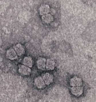
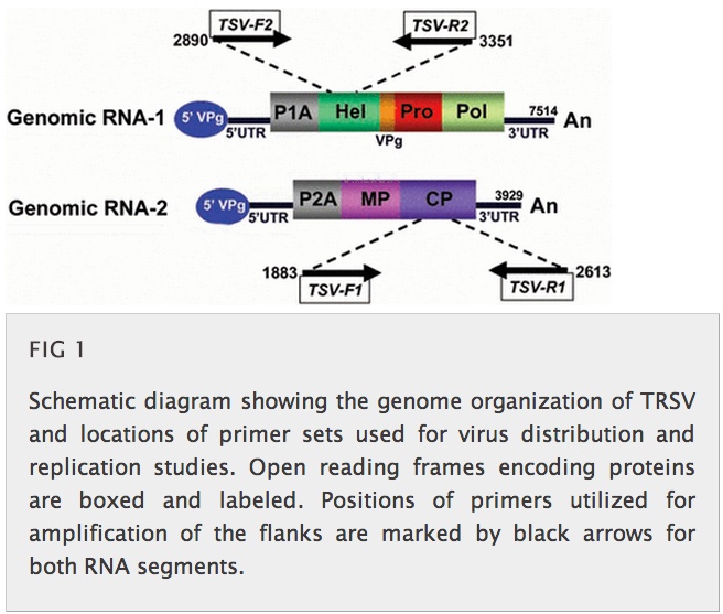
Another reservation I have concerns their methodology.  First, while I was impressed that they did strand-specific PCR to show  both the presence of viral (+ strand) and of replicative form (- strand) RNA in bee and mite tissue (see their Figure above), and did in situ hybridisation to show (-) strand presence in mites, they did not do something very simple that could have shown the same thing, AND given them genome-length +/- strand RNA to play with.

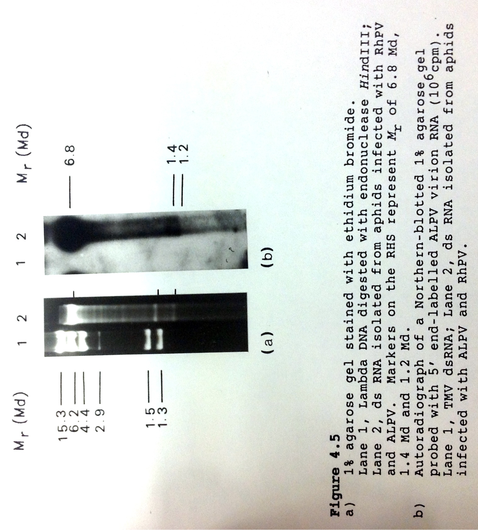
I refer, of course, to dsRNA isolation, which is a very easy and extremely clean technique that can be used to get full-length dsRNAs for many (+) strand RNA viruses from plant or insect tissues.  Moreover, the simple fact of isolating dsRNA forms for a single-strand RNA virus is indicative that replication is occurring – and was used by our group as long ago as 1988 (C Williamson, PhD Thesis, UCT) to isolate full-length ~10 kb dsRNA for two aphid picorna-like viruses.

Scan 04 Jan 2014, 16.31-page8

This means they could have had clear and simple evidence via dsRNA extraction of ALL of the coinfecting viruses present – without all of the expense of total cDNA sequencing.  And sampled more hives….

Second, I have to echo Adrian: “…no tests were done to show whether the virus isolated from bees infected known plant hosts of TRSV”.  Why ever not?  I am afraid that if I were a referee, I would have insisted on this: they did enough other work, after all, that this would not exactly have been an onerous requirement!

And here’s another thing: the authors say, in their Discussion,

“The finding from this study illustrates the complexity of relationships between plant pathogens and the pollinating insects and emphasizes the need for surveillance for potential host-jumping events as an integrated part of insect pollinator conservation.”

Ummmm…no, it doesn’t.  This is overstating the significance of their results by an order of magnitude at least.  They have simply illustrated that ONE species of honeybee may be infectable by ONE species of plant virus, and that this is ASSOCIATED with “weak” colonies.  Moreover, while the presence of TRSV was apparently associated with four weak colonies (out of only ten surveyed), it is quite possible that this is simply the emergence of a commensal-type infection against a background of known bee viruses, and in particular Israeli acute paralysis virus which was found in the same colonies (and blogged on here).  The authors also seem to take it as a given that the “emergence” of TRSV into bees is a recent jump – when it may not be recent at all.  Their statement in the abstract that

“The tree topology indicated that the TRSVs from arthropod hosts shared a common ancestor with those from plant hosts and subsequently evolved as a distinct lineage after transkingdom host alteration”

is pretty much unsubstantiated, in the absence of any investigation of the lineage in plants or in other bee colonies.  Further, they say that

“This study represents a unique example of viruses with host ranges spanning both the plant and animal kingdoms. “

Ummmmm….it doesn’t really do that, either: there are a LOT of arboviruses, with quite a few of them infecting insects and plants.  Here, for example, is an illustration from my teaching material of why it is that I think that viruses of insects and plants are an underappreciated evolutionary link for later evolution of viruses that got into mammals.

Transkingdom viruses

I note that bunyaviruses, rhabdoviruses, reoviruses and (not shown) picorna-like viruses appear linked by the fact that insects have possibly the most diverse representatives of these families, which may indicate that these originated in insects.  Which were the first complex animals to crawl out of the oceans, to join…plants on dry land?  Which explains how plants link up with the far more closely related (in evolutionary terms) insects and vertebrates: plants and insects were alone together for a long, long time before things with spines lurched up out of the water to join them.  So were their viruses.

I also said the following in the material there:

“A complicating factor in the picture of viruses co-evolving with their hosts over millennia is the fact that viruses apparently can – and obviously do – make big jumps in hosts every now and then.  It seems obvious, for example, that arthropods are almost certainly the original source for a number of virus families infecting insects and mammals – such as the Flaviviridae – and probably also of viruses infecting insects and other animals and plants – such as the Rhabdoviridae and Reoviridae – as well (see also here).  For example, picornaviruses of mammals are very similar structurally and genetically to a large number of small RNA viruses of insects and to at least two plant viruses, and – as the insect viruses are more diverse than the mammalian viruses – probably had their origin in some insect that adapted to feed on mammals (or plants) at some distant point in evolutionary time.”

Now quite a lot of interest has been shown in this paper in the blogosphere, and there have been quite a few conclusions drawn from the results that I think are largely unsubstantiated.  For example, this Sci Am blog claims

“When HIV jumped from chimpanzees to humans sometime in the early 1900s, it crossed a gulf spanning several million years of evolution. But tobacco ringspot virus, scientists announced last week, has made a jump that defies credulity. It has crossed a yawning chasm ~1.6 billion years wide.”

Again, ummmm…in light of the discussion above, not necessarily!  I am of the opinion that picorna-like viruses were shared between insects and plants, and then between insects and animals, hundreds of millions of years ago.  And TRSV is a nepovirus – and nepoviruses look like nothing more or less than a picornavirus with a divided genome.

I think TRSV represents something coming back into insects.  And I think we will probably find a lot more of them.

ViroBlogy: 2012 in review

1 February, 2013

So: thank you, anyone who clicked in, and regular visitors.  You make it worthwhile!!

The WordPress.com stats helper monkeys prepared a 2012 annual report for this blog.

Here’s an excerpt:

4,329 films were submitted to the 2012 Cannes Film Festival. This blog had 33,000 views in 2012. If each view were a film, this blog would power 8 Film Festivals

Click here to see the complete report.

Together, we can do more….

17 October, 2012

It gives me great delight to pass on some news about an old friend: I have co-authored two papers with the Pappus (husband and wife), and have maintained a long association with Hanu as a favoured referee for Archives of Virology; he has gone on to achieve some distinction at Washington State University – and recently to have made a fundamental discovery in plant virology.  I thank Eric Sorenson of the Washington State Magazine for sending me this.

Viral alliances overcome plant defenses, according to newly published WSU research

Contact:
Hanu Pappu, professor and chair of plant pathology, Washington State University, 509-335-3752, hrp@wsu.edu

PULLMAN, Wash. – Washington State University researchers have found that viruses will join forces to overcome a plant’s defenses and cause more severe infections.

“These findings have important implications in our ability to control these viruses,” says Hanu Pappu, Sam Smith Distinguished Professor of Plant Virology and chair of WSU’s Department of Plant Pathology. “Mixed infections are quite common in the field, and now we know that viruses in these mixed infections are helping each other at the genetic level to overcome host defenses and possibly lead to the generation of new viruses.”

Pappu publishes his findings in the latest issue of the journal PLOS ONE. Joining him are Ph.D. student Sudeep Bag and Neena Mitter, associate professor at Australia’s University of Queensland.

The researchers focused on iris yellow spot virus and tomato spotted wilt virus after Bag discovered that, when they infect the same plant, they helped each other overcome a plant’s defense response. With Mitter’s help and sophisticated molecular techniques, Bag found both viruses dramatically changed their genetic expression, breaking down the plant’s defenses and leading to more severe disease.

Bag also found that genes from the tomato spotted wilt virus seemed to “aid and abet” iris yellow spot virus as it spread throughout the plant and caused more disease.

Growers should take this phenomenon into account, says Pappu, with broader management tactics that target more than one virus and possible variations.

The research was funded in part by the Specialty Crops Research Initiative of the National Institute of Food and Agriculture, a branch of the U.S. Department of Agriculture.

The paper, “Complementation between Two Tospoviruses Facilitates the Systemic Movement of a Plant Virus Silencing Suppressor in an Otherwise Restrictive Host,” can be found athttp://dx.plos.org/10.1371/journal.pone.0044803.

PS: the Pappus cook REALLY good food – as I discovered in Florida, at Chuck Niblett’s house, back in 1996 or so….

White death: A diabolical pact between an insect and two viruses

22 August, 2012

See on Scoop.itVirology News

This is actually an article in The Economist from 2007 – forwarded to me by a Professor of Philosophy, as it happens, and which has mouldered on my desk lo, these past five years.  Thanks David Benatar!

“Whiteflies are pests in every continent that they are found in—and they are found in every continent except Antarctica. They cause damage directly, by consuming plant juices, and indirectly, by spreading viral diseases. But Liu Shusheng, of Zhejiang University, in Hangzhou, and his colleagues have found a strain of the species that delivers a double whammy. Not only does it spread diseases, but it is also vastly more successful when it lives on plants infected with the diseases in question [tomato yellow leafcurl and tobacco curly shoot begomoviruses, both ssDNA geminiviruses]  than when it subsists on healthy plants.”

This is a fascinating example of just why it is that certain vector-virus-host combinations can lead to success of the vector, and increased spread of the virus.  Basically,

“…type B insects lived six times longer on infected plants than uninfected ones, and their population per infected plant might rise as high as 13 times that on an uninfected one”

This means that geminivirus infection of host plants actually gives a survival advantage to the insects which transmit them.  Simple if unfortunate!!

See on www.economist.com