The very early discovery of avian viruses associated with cancer, and the subsequent failure for many years to isolate similar viruses from mammals, gave some researchers the idea that possibly birds were unique in this regard. However, “RNA tumour viruses” or oncornaviruses, as they were known for a time, were first demonstrated to affect mammals when mouse mammary tumours were shown to be due to a virus by John Bittner in 1936, by transmission in milk. He also demonstrated vertical transmission, or inheritance of the virus.
The nature of the agent was not known at the time, but by 1951 L Gross had shown that leukaemia could be passaged in mice using cell-free extracts. In 1958 W Bernhard had proposed a classification of what were to become known as retroviruses on the basis of electron microscopy. In 1964 a mouse sarcoma virus and a feline leukaemia virus had been isolated, and in 1969 bovine leukaemia was shown to be a viral disease. 1970 saw the description of reverse transcriptase from retroviruses, and in 1971 the first primate leukaemia virus – from gibbons – was described, and the first retrovirus (foamy virus) described from humans. Bovine leukaemia virus was characterised as a retrovirus in 1976.
It is not surprising, therefore, that many labs tried to find cancer-causing disease agents in humans. However, such effort had been put into finding oncornaviruses associated with human tumours, with such lack of success, that it led to people talking of “human rumour viruses” – a useful list of which can be seen here. Nevertheless, by 1980 Robert Gallo’s group had succeeded in finding “type C retrovirus particles from fresh and cultured lymphocytes of a patient with cutaneous T-cell lymphoma”, which they called human T-cell leukaemia virus (HTLV). The breakthrough was made possible by their prior discovery of “T cell growth factor”, now called interleukin 2 (IL-2), which meant human T cells could be successfully cultured for the first time. A group of Japanese researchers described an “Adult T cell leukemia virus” (ATLV) in 1982: this proved to be the same as what became HTLV-1, given the description also in 1982 by Gallo’s group of another retrovirus associated with a T-cell variant of hairy cell leukaemia, which they dubbed HTLV-2.
HTLV-1 is associated with the rare and genetically-linked adult T-cell leukaemia, found mainly in southern Japan, as well as with a demyelinating disease called “HTLV-I associated myelopathy/tropical spastic paraparesis (HAM/TSP)” and HTLV-associated uveitis and infective dermatitis. The areas of highest prevalence are Japan, Africa, the Caribbean islands and South America. HTLV-2 had a mainly Amerindian and African pygmy distribution, although it is now found worldwide, and causes a milder form of HAM/TSP, as well as arthritis, bronchitis, and pneumonia. It is is also frequent among injecting drug users. However, except for rare incidences of cutaneous lymphoma in people coinfected with HIV, and the fact of its origin in a hairy cell leukaemia, there is no good evidence that HTLV-2 causes lymphoproliferative disease. The two viruses infect between 15 and 20 million people worldwide. HTLV-1 infections can lead to an often rapidly fatal leukaemia.
By 2005 another two viruses had joined the family: HTLV-3 and HTLV-4 were described from samples from Cameroon that were presumably zoonoses – being associated with bushmeat hunters – and which are not associated with disease. Interestingly, all the HTLVs have simian counterparts – indicating species cross-over at some point in their evolution. Collectively they are known as the primate T-lymphotropic viruses (PTLVs) as they consitute an evolutionarily related group. Another relative is bovine leukaemia virus.
The HTLV-1/STLV-1 and HTLV-2/STLV-2 relationships are relatively ancient, at more than 20 000 years since divergence. However, their evolution differs markedly in that STLV-I occurs in Africa and Asia among at least 19 species of Old World primates, while STLV-2 has only been found in bonobos, or Pan paniscus dwarf chimpanzees from DR Congo. It is therefore quite possible that there are other HTLVs undiscovered in primates in Africa and elsewhere, that may yet emerge into the human population.
Human immunodeficiency virus type 1 (HIV-1) was for a time after its discovery in 1983 called HTLV-III by the Gallo group and lymphadenopathy virus (LAV) by the Montagnier group; however, evidence later obtained from sequencing and genome organisation showed by 1986 that it was in fact a lentivirus, related to viruses such as feline immunodeficiency virus (FIV) and the equine infectious anaemia virus discovered in 1904, and it was renamed. Francoise Barre-Sinoussie and Luc Montagnier were awarded a half share in a 2008 Nobel Prize, commemorated here
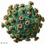
HIV particle. Russell Kightley Media
in Viroblogy.
HIV is indirectly implicated in cancer because it creates an environment through immunosuppression that allows the development of opportunistic tumours that would normally be controlled by the immune system: these include HPV-related cervical cancer, and Kaposi’s sarcoma caused by Human herpesvirus 8 (see later). It is also possible that HIV may directly cause lymphoma development in AIDS patients by insertional activation of cellular oncogenes, although this appears to be rare.


