+MaryMangan over there on Google+ made an interesting point about simple messages to refute the kinds of nonsense promulgated by vaccine denialists, among others.
Here’s my contribution:
Vaccines!
+MaryMangan over there on Google+ made an interesting point about simple messages to refute the kinds of nonsense promulgated by vaccine denialists, among others.
Here’s my contribution:
Vaccines!
See on Scoop.it – Virology and Bioinformatics from Virology.ca
ProMED Mail is possibly THE premier infectious disease updating service in the world today, having sprung to fame during the Kikwit Ebola outbreak in 1995. This is one of a series of posts on measles, which is documenting a very disturbing trend: the incidence of the disease is increasing in places where it should have been eradicated, because well-educated and sophisticated communities are not vaccinating their children – or themselves.
One very telling quote from the post:
“Measles is highly infectious so we must all do
everything possible to prevent the spread of it, particularly with an outbreak on our doorstep. … MMR vaccination is the only way to prevent
measles. If parents haven’t arranged for their children to be vaccinated – it’s not too late to have the jab. Parents don’t realise that measles is not just a case of a few spots – it can be a very serious illness. Symptoms include fever, cough, soreness of the eyes and a rash which spreads rapidly over the body. Serious complications affect one in 15 children. These include chest infections, fits, encephalitis (swelling of the brain), and brain damage. In very serious cases, measles can kill.”
See on www.promedmail.org
A technical development that was to greatly advance the study of viruses was begun in 1923, but only reached fruition by the 1930s: this was the ultracentrifuge, invented and developed first by Theodor (“The”) Svedberg in Sweden as a purely analytical tool, and later by JW Beams and EG Pickels in the USA as an analytical and preparative tool. The ultracentrifuge revolutionised first, the physical analysis of proteins in solution, and second, the purification of proteins, viruses and cell components, by allowing centrifugation at speeds high enough to allow pelleting of subcellular fractions.
Analytical centrifugation and calculation of molecular weights of particles gave some of the first firm evidence that certain proteins, and virus particles, were large, regular objects. Indeed, it came to be taken as a given that one of the fundamental properties of a virus particle was its sedimentation coefficient, measured in svedbergs (a unit of 10-13 seconds, shown as S20,W). This is also how ribosomes of pro- and eukaryotes came to be named: these are known as 70S (prokaryote) and 80S ribosomes, respectively, based on their different sedimentation rates.
In 1931, Robert Shope in the USA managed to recreate swine influenza by intranasal administration of filtered secretions from infected pigs. Moreover, he showed that the classic severe disease required co-inoculation with a bacterium – Haemophilus influenza suis – originally thought to be the only agent. He also pointed out the similarities between the swine disease and the Spanish Flu, where most patients died of secondary infections. However, he also suggested that the virus survived seasonally in a cycle involving the pig, lungworms, and the earthworm, which is now known to be completely wrong.
This notwithstanding, he found that people who had survived infection during the 1918 pandemic had antibodies protecting them against the swine flu virus, while people born after 1920 did not, which showed that the 1918 human and swine flu viruses were very similar if not identical. This was a very relevant discovery for what happened much later, in the 2009 influenza pandemic, when the same virus apparently came back into the human population from pigs after circulating in them continuously since 1918.
Shope went on in 1932 to discover, with Peyton Rous, what was first called the Shope papillomavirus and later Cottontail rabbit papillomavirus: this causes benign cancers in the form of long hornlike growths on the head and face of the animal. This may explain the sightings in the US Southwest of the near-mythical “jackalope”.
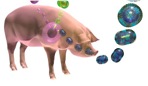
Influenza viruses in pigs
Patrick Laidlaw and William Dunkin, working in the UK at the National Institute for Medical Research (NIMR), had by 1929 successfully characterised the agent of canine distemper – a relative of measles, mumps and distemper morbilliviruses – as a virus, proved it infected dogs and ferrets, and in 1931 got a vaccine into production that protected dogs. This was made from chemically inactivated filtered tissue extract from infected animals. Their work built on and completely eclipsed earlier findings, such as those of Henri Carré in France in 1905, who first claimed to have shown it was a filterable agent, and Vittorio Puntoni, who first made a vaccine in Italy from virus-infected brain tissue inactivated with formalin in 1923.
Continuing from Laidlaw and Dunkin’s work in the same institute, Christopher Andrewes, Laidlaw and W Smith reported in 1933 that they had isolated a virus from humans infected with influenza from an epidemic then raging. They had done this by infecting ferrets with filtered extracts from infected humans – after the fortuitous observation that ferrets could apparently catch influenza from infected investigators! The “ferret model” was very valuable – see here for modern use of ferrets – as strains of influenza virus could be clinically distinguished from one another.

Influenza virus and eggs: large-scale culture
Frank Macfarlane Burnet from Australia visited the NIMR in the early 1930s, and learned a number of techniques he used to great effect later on. Principal among these was the technique of embryonated egg culture of viruses – which he took back to Melbourne, and applied to the infectious laryngotracheitis virus of chickens in 1936. This is a herpesvirus, first cultivated by JR Beach in the USA in 1932: Burnet used it to demonstrate that it was possible to do “pock assays” on chorioallantoic membranes that were very similar to the plaque assays done for bacteriophages, with which he was also very familiar. Also in 1936, Burnet started a series of experiments on culturing human influenza virus in eggs: he quickly showed that it was possible to do pock assays for influenza virus, and that
“It can probably be claimed that, excluding the bacteriophages, egg passage influenza virus can be titrated with greater accuracy than any other virus.”
Max Theiler and colleagues in the USA took advantage of the new method of egg culture to adapt the French strain of yellow fever virus (YFV) he had grown in mouse brains to being grown in chick embryos, and showed that he could attenuate the already weakened strain even further – but it remained “neurovirulent”, as it caused encephalitis or brain inflammation in monkeys. He then adapted the first YFV characterised – the Asibi strain, from Ghana in 1927 – to being grown in minced chicken embryos lacking a spinal cord and brain, and showed in 1937 that after more than 89 passages, the virus was no longer “neurotrophic”, and did not cause encephalitis. The new 17D strain of YFV was successfully tested in clinical trials in Brazil in 1938 under the auspices of the Rockefeller Foundation, which has supported YFV work since the 1920s. The strain remains in use today, and is still made in eggs.
Given that the nature of viruses had prompted people to think of them as “chemical matter”, researchers had attempted from early days to isolate, purify and characterise the infectious agents. An early achievement was the purification of a poxvirus in 1922 by FO MacCallum and EH Oppenheimer.
Much early work was done with bacteriophages and plant viruses, as these were far easier to purify or extract at the concentrations required for analysis, than animal or especially human viruses.
CG Vinson and AM Petre, working with the infectious agent causing mosaic disease in tobacco – tobacco mosaic virus, or TMV – showed in 1931 that they could precipitate the virus from suspension as if it were an enzyme, and that infectivity of the precipitated preparation was preserved. Indeed, in their words:
“…it is probable that the virus which we have investigated reacted as a chemical substance”.
Viruses in Crystal
An important set of discoveries started in 1935, when Wendell Stanley in the USA published the first proof that TMV could be crystallised, at the time the most stringent way of purifying molecules. He also reported that the “protein crystals” were contaminated with small amounts of phosphorus. An important finding too, using physical techniques including ultracentrifugation and later, electron microscopy, was that the TMV “protein” had a very high molecular weight, and was in fact composed of large, regular particles. This was a very significant discovery, as it indicated that some viruses at least really were very simple infectious agents indeed.

TMV particle: 95% protein, 5% RNA
However, his conclusion that TMV was composed only of protein was soon challenged, when Norman Pirie and Frederick Bawden working in the UK showed in 1937 that ribonucleic acid (RNA) – which consists of ribose sugar molecules linked by phosphate groups – could be isolated consistently from crystallised TMV as well as from a number of other plant viruses, which accounted for the phosphorus “contamination”. This resulted in the realisation that TMV and other plant virus particles – now known to be virions – were in fact nucleoproteins, or protein associated with nucleic acid.
Stanley received a share of the Nobel Prize in Chemistry in 1946 for his work on TMV: it is instructive to read his acceptance speech from the time to realise what the state of the science that was becoming virology was at the time. He wrote:
“Since the original discovery of this infectious, disease-producing agent, known as tobacco mosaic virus, well over three hundred different viruses capable of causing disease in man, animals and plants have been discovered. Among the virus-induced diseases of man are smallpox, yellow fever, dengue fever, poliomyelitis, certain types of encephalitis, measles, mumps, influenza, virus pneumonia and the common cold. Virus diseases of animals include hog cholera, cattle plague, foot-and-mouth disease of cattle, swamp fever of horses, equine encephalitis, rabies, fowl pox, Newcastle disease of chickens, fowl paralysis, and certain benign as well as malignant tumors of rabbits and mice. Plant virus diseases include tobacco mosaic, peach yellows, aster yellows, potato yellow dwarf, alfalfa mosaic, curly top of sugar beets, tomato spotted wilt, tomato bushy stunt, corn mosaic, cucumber mosaic, and sugar cane yellow stripe. Bacteriophages, which are agents capable of causing the lysis of bacteria, are now regarded as viruses”.
Two of the most interesting things about the article, however, are the electron micrographs of virus particles – Stanley had one of the first electron micrsoscopes available at the time – and the table of sizes of viruses, proteins and cells that had been determined by then by techniques such as ultracentrifugation and filtration: TMV was known to be rodlike, 15 x 280 nm; vaccinia was 210 x 260 nm; poliomyelitis was 25 nm; phages like T2 were known to have a head-and-tail structure.
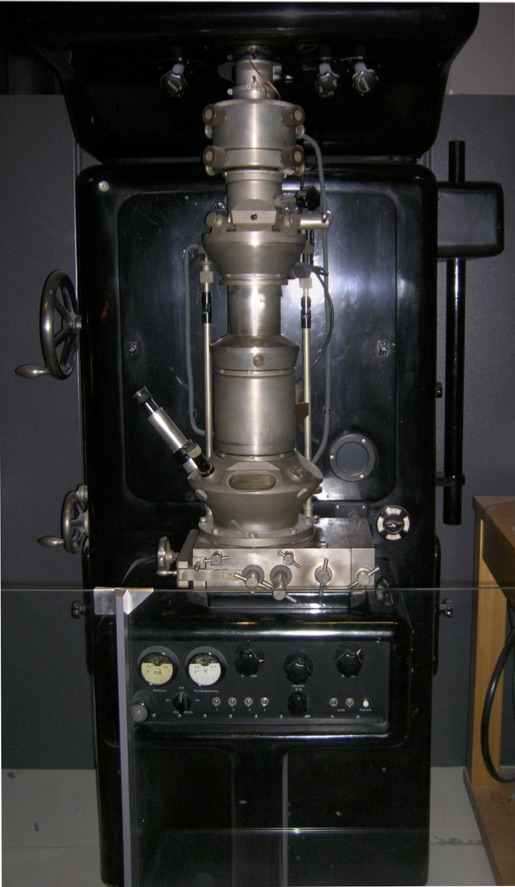
First Electron Microscope with Resolving Power Higher than that of a Light Microscope. Ernst Ruska, Berlin 1933
Wikipedia CC BY-SA 3.0, https://www.flickr.com/photos/93452909@N00/176059674
The development of the electron microscope, in Germany in the 1930s, represented a revolution in the investigation of virus structures: while virions of viruses like variola and vaccinia could just about be seen by light microscopy – and had been, as early as 1887 by John Buist and others – most viruses were far too small to be visualised in this way.
While Ernst Ruska received a Nobel Prize in 1986 for developing the electron microscope, it was his brother Helmut who first imaged virus particles – using beams of electrons deflected off virus particles coated in heavy metal atoms. From 1938 through the early 1940s, using his “supermicroscope”, he imaged virions of poxviruses, TMV, varicella-zoster herpesvirus, and bacteriophages, and showed that they were all particulate – that is, they consisted of regular and sometimes complex particles, and were often very different from one another. He even proposed in 1943 a system of viral classification on the basis of their perceived structure.
While electron microscopy was also used medically to some extent thereafter – for example, in differentiating smallpox from chickenpox by imaging particles of variola virus and varicella-zoster virus, respectively, derived from patients’ vesicles – its use was limited by the expense and cumbersome nature of sample preparation. For example, the micrographs in Stanley’s 1946 paper were all done with samples “…prepared with gold by the shadow-casting technique”.
The use of the cumbersome technique of metal shadow-casting, and the highly inconvenient nature of electron microscopy as a routine tool all changed from 1959 onwards, when Sydney Brenner and Robert Horne published “A negative staining method for high resolution electron microscopy of viruses”. This method involves the use of viruses in liquid samples deposited on carbon-coated metal grids, and then stained with heavy-metal salts such as phosphotungstic acid (PTA) or uranyl acetate.
This simple technique revolutionised the field of electron microscopy, and within just a few years much information was acquired about the architecture of virus particles. Not only were the overall shapes of particles revealed, but also the details of the symmetrical arrangement of their components. Some beautiful examples can be seen here, at the Cold Spring Harbor site.

Depiction of the effects of using a heavy metal salt solution to negatively stain particles on a carbon film. The stain (dark) pools around the particles (light). Human rotavirus particles, stained from below (left) and by immersion (right). Images copyright LM Stannard
Click here for Part 1: Filters and Discovery
here for Part 3: Phages, Cell Culture and Polio
and here for Part 4: RNA Genomes and Modern Virology
Copyright Edward P Rybicki and Russell Kightley, February 2015, except where otherwise noted.
At last, someone heavy has gone and nailed it down: The Scientist (http://the-scientist.com/2011/08/26/vaccines-are-safe/) reports that
“Vaccines are safe and not the cause of autism, according to a new report from the Institute of Medicine, the health arm of the National Academies. The panel based its conclusions on the review of more than 1,000 studies on eight vaccines commonly given to children, including those for chickenpox, meningitis, tetanus, and measles, mumps, and rubella (MMR).”
One. Thousand. Studies. At least! There’s more:
“Some serious side effects were linked to vaccines, but occurred very rarely. Among them: those who receive the chicken pox vaccine could later come down with pneumonia or meningitis if their immune systems become compromised by diseases such as cancer, and the MMR vaccine occasionally sets off brain inflammation or seizures, Nature reports. Six of the eight vaccines can also cause allergic reactions. The more serious side effects most commonly occur in children who have underlying immune problems.”
As I taught my captive second- and third-years for close on thirty years, there’s a risk-benefit calculation to be done for every vaccine, and indeed, for every drug that you or yours may be exposed to. If there’s no benefit – because there’s no risk – then do without. If, on the other hand, your baby stands a reasonable chance of getting seriously ill, and maybe even suffering permanent damage from getting infected by a preventable dissease – vaccinate!
And that’s rotavirus I’m talking about, not even something really nasty like measles. I did the same survey over several years, asking 70+ students if they would give kids a vaccine that had a POSSIBLE 1/40 000 chance of causing a possibly fatal intestinal complication (intussusseption, or telescoping of the bowel) in (a) an environment where no children died of rotavirus, (b) an environment like in many places in the developing world, where 1/100 children might die. Nearly every single one, every time, would not use it in (a), but would have no hesitation in scenario (b).
Regulars on this blog will know what I think about not vaccinating against measles. So now, folks: reach out to a relative or a friend who espouses this utter nonsense linking vaccines to autism – and smack them solidly. Then get their kids vaccinated.
OK, maybe not – but remonstrate with them, point them towards the evidence, tell them just how nasty some of the preventable diseases can be, and that they should realise measles, polio or rotavirus are only someone else’s plane trip away.
I have written previously about the Rinderpest virus eradication campaign – and now it appears as though the final nail has in fact been hammered into the coffin’s lid while I wasn’t looking. I thank my son Steven for noticing!
It was officially announced on 26th June, in Rome – the headquarters of the Food and Agriculture Organisation (FAO) – that “…for only the second time in history, a disease has been wiped off the face of the earth”.
From a New York Times article:
The long but little-known campaign to conquer rinderpest is a tribute to the skill and bravery of “big animal” veterinarians, who fought the disease in remote and sometimes war-torn areas — across arid stretches of Africa bigger than Europe, in the Arabian desert and on the Mongolian steppes.
…
The victory is also proof that the conquest of smallpox was not just an unrepeatable fluke, a golden medical moment that will never be seen again. Since it was declared eradicated in 1980, several other diseases — like polio, Guinea worm, river blindness, elephantiasis, measles and iodine deficiency — have frustrated intensive, costly efforts to do the same to them. The eradication of rinderpest shows what can be done when field commanders combine scientific advances and new tactics.… The modern eradication campaign began in 1945, when the Food and Agriculture Organization was founded. But it became feasible only as vaccines improved. An 1893 version made from the bile of convalescent animals was replaced by vaccines grown in goats and rabbits and finally in laboratory cell lines; a heat-stable version was developed in the 1980s.
The interesting thing about Rinderpest virus is that it is probably a consequence of human’s development of agriculture and especially the keeping of livestock, that got it into animals in the first place: it is closely enough related to measles virus that it probably only diverged from it some time around CE 1000.
So it’s not just us that get animal viruses – our pets and our livestock can get them from us, too.
But it’s now time to concentrate on the next two: polioviruses and measles. Vaccinate, brothers and sisters, vaccinate!!
I have written before in ViroBlogy about measles resurging in Africa (see: Measles in Zimbabwe from January 2010) – and now Larry Madoff, the Editor of the very worthy ProMED, makes the case that it is resurging all over. And in the case of developed countries, largely because of simple stupidity.
From Larry:
Once nearly eradicated in much of the developed world, measles outbreaks are becoming more frequent in 2011. They are the result of increased global travel, lower rates of vaccination in poorer counties – and parents choosing not to vaccinate their children in the U.S., Europe and elsewhere [my emphasis] because of the now widely discredited myth that the measles, mumps and rubella vaccine causes autism.
Whenever people are on the move, there are risks of infectious diseases moving with them.
… [ fundraising message removed: go here to donate]
Measles kills an estimated 165,000 people each year, mostly in poor countries. Since January, however, measles outbreaks reported on ProMED mail have occurred not only in poorer nations such as Bangladesh, Somalia, and Pakistan, but in such countries as France, Spain, England, Canada, Australia, New Zealand, and within the U.S. from Massachusetts to Utah, Minnesota to New Jersey. An outbreak of measles in the Canary Islands and several South American countries, in fact, appears to be the result of unvaccinated British and German tourists bringing the disease to their shores.
As I said, then: stupidity, in the case of unvaccinated tourists. And lack of vaccine or problems in delivery in the case of the poorer nations.
The first is easy to fix: simply don’t let any tourists in without proof of measles vaccination, as presently happens in Brazil for yellow fever, for example. It would be done for all the wrong reasons, but hey, whatever works!
The second…is harder. Measles vaccines are good: they are effective and safe, whether given singly or in combination (eg: measles-mumps-rubella; MMR) – and pretty cheap; cheap enough to be included in the free Extended Programme of Immunisation (EPI) bundle in many countries. But the simple fact is that they are not getting to many of the folk who need them – and given that you need a minimum of 80% coverage to get “herd immunity”, the virus just keeps on being transmitted around.
And measles is not a trivial disease, whatever the lay population thinks: if it can kill or cause severe complications in healthy, well-fed children, imagine how much worse the consequences of infection are in malnourished, sickly children. As mentioned above, 165 000 people – and mainly children – die every year from measles.
From the WHO Measles Fact Sheet:
Measles is a highly contagious vaccine-preventable disease caused by the measles virus, a member of the genus Morbillivirus in the family Paramyxoviridae. It is spread by droplets or direct contact with nasal or throat secretions of infected persons; less commonly by airborne spread or by articles freshly soiled with secretions of nose and throat. Measles is one of the most readily transmitted communicable diseases and probably the best known and most deadly of all childhood rash/fever illnesses. [my emphasis].
The scale of the problem can be seen here:

Anywhere that isn’t blue has less than 90% coverage – and look at Africa…mostly 50-79% coverage, and that is simply not enough.
Look again at the complications of natural measles infections – from the CDC Measles Complications page:
About 30% of measles cases develop one or more complications, including
- Pneumonia, which is the complication that is most often the cause of death in young children.
- Ear infections occur in about 1 in 10 measles cases and permanent loss of hearing can result.
- Diarrhea is reported in about 8% of cases.
These complications are more common among children under 5 years of age and adults over 20 years old.
Even in previously healthy children, measles can be a serious illness requiring hospitalization. As many as 1 out of every 20 children with measles gets pneumonia, and about 1 child in every 1,000 who get measles will develop encephalitis. (This is an inflammation of the brain that can lead to convulsions, and can leave the child deaf or mentally retarded.) For every 1,000 children who get measles, 1 or 2 will die from it. Measles also can make a pregnant woman have a miscarriage, give birth prematurely, or have a low-birth-weight baby.
In developing countries, where malnutrition and vitamin A deficiency are common, measles has been known to kill as many as one out of four people. It is the leading cause of blindness among African children. [my emphases]
Sub-acute sclerosing panencephalitis (SSPE) is a very rare, but fatal degenerative disease of the central nervous system that results from a measles virus infection acquired earlier in life. Analysis of data from an outbreak of measles in the United States during 1989-1991 suggests a rate of 4-11 cases of SSPE per 100,000 cases of measles. A risk factor for developing this disease is measles infection at an early age.
If this doesn’t scare you, then you are invincibly ignorant. Or simply stupid. Which the Bill & Melinda Gates Foundation is not – they have supported new measles vaccine development since 2000; they are also recently involved along with the Lions Club International Foundation in the Measles Initiative, which is:
“…a worldwide effort to protect children from measles and strengthen routine immunization services. UNICEF, World Health Organization (WHO), U.S. Centers for Disease Control (CDC), American Red Cross, and the United Nations Foundation are among the organizations contributing to these efforts since 2001.”
From their site:
An estimated 164,000 people – 450 a day – died from this easily preventable disease in 2008. Costing less than US $1 to vaccinate a child, the measles control strategy represents one of the most cost-effective health interventions available.
Yet, many developing countries that are facing multiple health challenges have limited funds, making financial support from the Measles Initiative critical. A steep decline in donor investment has resulted in a significant funding gap. Unless conditions improve, the shortfall will put the goal and millions of children at risk.
We can eradicate measles. We really, really can – but it starts with vaccinating your children, and yourself. Then helping vaccinate others.
Rinderpest virus infects cattle, buffalo and several species of antelope among other animals: it is a member of the genus Morbillivirus,family Paramyxoviridae, and is related to measles and mumps viruses in humans, distemper virus in dogs, and a variety of relatively newly-described viruses in marine mammals. It also almost certainly gave rise to measles virus sometime around the 11th-12th centuries CE, as an originally zoonotic infection – sourced in domestic animals – took root in humans and began to be passed around (see MicrobiologyBytes).

Electron micrograph of a morbillivirus particle showing the membrane, matrix, and inner helical nucleocapsid. Image by LM Stannard
The ICTVdB generic description of morbilliviruses is as follows:
Virions consist of an envelope and a nucleocapsid. Virus capsid is enveloped. Virions are spherical to pleomorphic; filamentous and other forms are common. Virions measure (60-)150-250(-300) nm in diameter; 1000-10000 nm in length. Surface projections are distinctive spikes of haemagglutinin (H) and fusion (F) glycoproteins covering evenly the surface. Surface projections are 9-15 nm long; spaced 7-10 nm apart. Capsid/nucleocapsid is elongated with helical symmetry. The nucleocapsid is filamentous with a length of 600-800(-1000) nm and a width of 18 nm. Pitch of helix is 5.5 nm.
The Mr of the genome constitutes 0.5% of the virion by weight. The genome is not segmented and contains a single molecule of linear negative-sense, single-stranded RNA. Virions may also contain occasionally a positive sense single-stranded copy of the genome (thus, partial self-annealing of extracted RNA may occur). The complete genome is 15200-15900 nucleotides long.
Wikipedia describes rinderpest virus as “…an infectious viral disease of cattle, domestic buffalo, and some species of wildlife. The disease was characterized by fever, oral erosions, diarrhea, lymphoid necrosis, and high mortality.” And: “The term Rinderpest is taken from German, and means cattle-plague.”
The Food and Agriculture Organisation (FAO) has a Division of Animal Production and Health: their web site details a campaign known as the Global Rinderpest Eradication Programme (GREP), which has been going since 1994.
With very little fanfare, I might point out: as a practicing teaching virologist, I was totally unaware of it. Anyway: they state that:
Rinderpest has been a dreaded cattle disease for millennia, causing massive losses to livestock and wildlife on three continents. This deadly cattle plague triggered several famines and caused the loss of draught animal power in agricultural communities in the 18th, 19th and 20th centuries.
…which is a little of an understatement: Wikipedia tells us that
“Cattle plagues recurred throughout history, often accompanying wars and military campaigns. They hit Europe especially hard in the 18th century, with three long pandemics which, although varying in intensity and duration from region to region, took place in the periods of 1709–1720, 1742–1760, and 1768–1786. There was a major outbreak covering the whole of Britain in 1865/66.”
“Later in history, an outbreak in the 1890s killed 80 to 90 percent of all cattle in Southern Africa, as well as in the Horn of Africa [and resulted in the deaths of many thousands of people who depended on them]. Sir Arnold Theiler was instrumental in developing a vaccine that curbed the epidemic. [my insert / emphasis] More recently, a rinderpest outbreak that raged across much of Africa in 1982–1984 cost at least an estimated US$500 million in stock losses”.
When commenting on the significance of the achievement, John Anderson, the head of the FAO, described GREP’s announcement that Rinderpest had been eradicated as:
The 19th century southern African outbreak was devastating enough that people still remember it as a legendary time of hardship – and then there was the 1980s outbreak. Another South African interest in rinderpest is that the legendary Sir Arnold Theiler had a hand in making a vaccine: he did this around the turn of the 20th century, by simultaneously injecting animals with blood from an infected animal and antiserum from a recovered animal: this protected animals for long enough to allow their immune systems to respond to the virus – but was rather risky, even though it was used for several decades.
In the 1920s J. T. Edwards in what is now the Indian Veterinary Research Institute serially passaged the virus in goats: after 600 passages it no longer caused disease, but elicited lifelong immunity. However, it could still cause disease in immunosuppressed cattle.
In 1962, Walter Plowright and R.D. Ferris used tissue culture to develop a live-attenuated vaccine grown in calf kidney cells. Virus that had been passaged 90 times conferred immunity without disease even in immunosuppressed cattle, was stable, and did not spread between animals. This vaccine was the one that allowed the prospect of eradicating the virus, and earned Plowright a World Food Prize in 1999.
But a memory may be all rinderpest is any more – as the GREP site says the following:
“The last known rinderpest outbreak in the world was reported in 2001 (Kenya). Based on the above-mentioned investigations, FAO is confident that all rinderpest virus lineages will prove to be extinct.”
This was also announced via the BBC on the 14th October, 2010. They said:
The eradication of the virus has been described as the biggest achievement in veterinary history and one which will save the lives and livelihoods of millions of the poorest people in the world.
And the significant bit:
If confirmed, rinderpest would become only the second viral disease – after smallpox – to have been eliminated by humans.
Let us reiterate that: only the second viral disease, ever, to have been eliminated. And how was this possible? Unlike smallpox, which has only humans as a natural and reservoir host (although it almost certainly also got into us from animals), rinderpest attacked a wider range of hosts. However, it seemed mainly to have a reservoir in domesticated cattle, and it did not have an arthropod vector; moreover, the vaccine was cheap and effective.
This is momentous news: we may well have succeeded in ridding the planet of what has been a very significant disease of livestock and of wild animals, which has caused untold agricultural loss throughout recorded history, and which has resulted in enormous human hardship as well. We have also made a natural species go extinct – but it won’t be missed. Like smallpox, it was completely sequenced some time ago, so we could theoretically recreate it if we ever needed to.
From GREP:
Though the effort to eradicate rinderpest has encountered many obstacles over the past several decades, the disease remains undetected in the field since 2001. As of mid 2010, FAO is confident that the rinderpest virus has been eliminated from Europe, Asia, Middle East, Arabian Peninsula, and Africa. This has been a remarkable achievement for veterinary science, evidence of the commitment of numerous countries, and a victory for the international community.
Amen. However – it’s not quite time to celebrate as the certification is only planned for 2011.
And now for mumps, and measles too.

