Posts Tagged ‘adenovirus’

Vaccination with Adenovirus Serotypes 35, 26, and 48 Elicits Higher Levels of Innate Cytokine Responses than Adenovirus Serotype 5 in Rhesus Monkeys

24 August, 2012

See on Scoop.itVirology and Bioinformatics from Virology.ca

“These data demonstrate that Ad35, Ad26, and Ad48, which utilize CD46 as their primary cellular receptor, induce significantly greater innate cytokine responses than Ad5, which uses the coxsackievirus and adenovirus receptor (CAR). These differences in innate triggering result in markedly different immunologic milieus for the subsequent generation of adaptive immune responses by these vaccine vectors.”


Important news for the vectored vaccine community in general, and for HIV vaccine in particular: Ad5 was the vehicle of choice; now it looks as though it shouldn’t be.


Adenovirus graphic courtesy of Russell Kightley Media

See on jvi.asm.org

A Short History of the Discovery of Viruses – Part 2

7 February, 2012

The following text has now appeared in modified form in an ebook, for sale for US$4.99 on the iBooks Store

The Ultracentrifuge, Eggs and Flu

The ultracentrifuge

A technical development that was to greatly advance the study of viruses was begun in 1923, but only reached fruition by the 1930s: this was the ultracentrifuge, invented and developed first by Theodor (“The”) Svedberg in Sweden as a purely analytical tool, and later by JW Beams and EG Pickels in the USA as an analytical and preparative tool.  The ultracentrifuge revolutionised first, the physical analysis of proteins in solution, and second, the purification of proteins, viruses and cell components, by allowing centrifugation at speeds high enough to allow pelleting of subcellular fractions.

Analytical centrifugation and calculation of molecular weights of particles gave some of the first firm evidence that certain proteins, and virus particles, were large, regular objects.  Indeed, it came to be taken as a given that one of the fundamental properties of a virus particle was its sedimentation coefficient, measured in svedbergs (a unit of 10-13 seconds, shown as S20,W).  This is also how ribosomes of pro- and eukaryotes came to be named: these are known as 70S (prokaryote) and 80S ribosomes, respectively, based on their different sedimentation rates.

The Official Discovery of Influenza Virus

In 1931, Robert Shope in the USA managed to recreate swine influenza by intranasal administration of filtered secretions from infected pigs.  Moreover, he showed that the classic severe disease required co-inoculation with a bacterium – Haemophilus influenza suis – originally thought to be the only agent.  He also pointed out the similarities between the swine disease and the Spanish Flu, where most patients died of secondary infections.  However, he also suggested that the virus survived seasonally in a cycle involving the pig, lungworms, and the earthworm, which is now known to be completely wrong.

This notwithstanding, he found that people who had survived infection during the 1918 pandemic had antibodies protecting them against the swine flu virus, while people born after 1920 did not, which showed that the 1918 human and swine flu viruses were very similar if not identical. This was a very relevant discovery for what happened much later, in the 2009 influenza pandemic, when the same virus apparently came back into the human population from pigs after circulating in them continuously since 1918.

Shope went on in 1932 to discover, with Peyton Rous, what was first called the Shope papillomavirus and later Cottontail rabbit papillomavirus: this causes benign cancers in the form of long hornlike growths on the head and face of the animal. This may explain the sightings in the US Southwest of the near-mythical “jackalope”.

Influenza viruses in pigs

Influenza viruses in pigs

Patrick Laidlaw and William Dunkin, working in the UK at the National Institute for Medical Research (NIMR), had by 1929 successfully characterised the agent of canine distemper – a relative of measles, mumps and distemper morbilliviruses – as a virus, proved it infected dogs and ferrets, and in 1931 got a vaccine into production that protected dogs.  This was made from chemically inactivated filtered tissue extract from infected animals.  Their work built on and completely eclipsed earlier findings, such as those of Henri Carré in France in 1905, who first claimed to have shown it was a filterable agent, and Vittorio Puntoni, who first made a vaccine in Italy from virus-infected brain tissue inactivated with formalin in 1923.

Influenza and Ferrets: the Early Days

Continuing from Laidlaw and Dunkin’s work in the same institute, Christopher Andrewes, Laidlaw and W Smith reported in 1933 that they had isolated a virus from humans infected with influenza from an epidemic then raging.  They had done this by infecting ferrets with filtered extracts from infected humans – after the fortuitous observation that ferrets could apparently catch influenza from infected investigators!  The “ferret model” was very valuable – see here for modern use of ferrets – as strains of influenza virus could be clinically distinguished from one another.

Eggs and Flu and Yellow Fever

Influenza virus and eggs: large-scale culture

Frank Macfarlane Burnet from Australia visited the NIMR in the early 1930s, and learned a number of techniques he used to great effect later on.  Principal among these was the technique of embryonated egg culture of viruses – which he took back to Melbourne, and applied to the infectious laryngotracheitis virus of chickens in 1936.  This is a herpesvirus, first cultivated by JR Beach in the USA in 1932: Burnet used it to demonstrate that it was possible to do “pock assays” on chorioallantoic membranes that were very similar to the plaque assays done for bacteriophages, with which he was also very familiar.  Also in 1936, Burnet started a series of experiments on culturing human influenza virus in eggs: he quickly showed that it was possible to do pock assays for influenza virus, and that

“It can probably be claimed that, excluding the bacteriophages, egg passage influenza virus can be titrated with greater accuracy than any other virus.”

Max Theiler and colleagues in the USA took advantage of the new method of egg culture to adapt the French strain of yellow fever virus (YFV) he had grown in mouse brains to being grown in chick embryos, and showed that he could attenuate the already weakened strain even further – but it remained “neurovirulent”, as it caused encephalitis or brain inflammation in monkeys.  He then adapted the first YFV characterised – the Asibi strain, from Ghana in 1927 – to being grown in minced chicken embryos lacking a spinal cord and brain, and showed in 1937 that after more than 89 passages, the virus was no longer “neurotrophic”, and did not cause encephalitis.   The new 17D strain of YFV was successfully tested in clinical trials in Brazil in 1938 under the auspices of the Rockefeller Foundation, which has supported YFV work since the 1920s.  The strain remains in use today, and is still made in eggs.

Virus purification and the physicochemical era

Given that the nature of viruses had prompted people to think of them as “chemical matter”, researchers had attempted from early days to isolate, purify and characterise the infectious agents.  An early achievement was the purification of a poxvirus in 1922 by FO MacCallum and EH Oppenheimer. 

Much early work was done with bacteriophages and plant viruses, as these were far easier to purify or extract at the concentrations required for analysis, than animal or especially human viruses. 

CG Vinson and AM Petre, working with the infectious agent causing mosaic disease in tobacco – tobacco mosaic virus, or TMV – showed in 1931 that they could precipitate the virus from suspension as if it were an enzyme, and that infectivity of the precipitated preparation was preserved.  Indeed, in their words:

“…it is probable that the virus which we have investigated reacted as a chemical substance”.

Viruses in Crystal

An important set of discoveries started in 1935, when Wendell Stanley in the USA published the first proof that TMV could be crystallised, at the time the most stringent way of purifying molecules.  He also reported that the “protein crystals” were contaminated with small amounts of phosphorus.  An important finding too, using physical techniques including ultracentrifugation and later, electron microscopy, was that the TMV “protein” had a very high molecular weight, and was in fact composed of large, regular particles.  This was a very significant discovery, as it indicated that some viruses at least really were very simple infectious agents indeed.

TMV particle: 95% protein, 5% RNA

However, his conclusion that TMV was composed only of protein was soon challenged, when Norman Pirie and Frederick Bawden working in the UK showed in 1937 that ribonucleic acid (RNA) – which consists of ribose sugar molecules linked by phosphate groups – could be isolated consistently from crystallised TMV as well as from a number of other plant viruses, which accounted for the phosphorus “contamination”.  This resulted in the realisation that TMV and other plant virus particles – now known to be virions – were in fact nucleoproteins, or protein associated with nucleic acid.

Stanley received a share of the Nobel Prize in Chemistry in 1946 for his work on TMV: it is instructive to read his acceptance speech from the time to realise what the state of the science that was becoming virology was at the time.  He wrote:

“Since the original discovery of this infectious, disease-producing agent, known as tobacco mosaic virus, well over three hundred different viruses capable of causing disease in man, animals and plants have been discovered. Among the virus-induced diseases of man are smallpox, yellow fever, dengue fever, poliomyelitis, certain types of encephalitis, measles, mumps, influenza, virus pneumonia and the common cold. Virus diseases of animals include hog cholera, cattle plague, foot-and-mouth disease of cattle, swamp fever of horses, equine encephalitis, rabies, fowl pox, Newcastle disease of chickens, fowl paralysis, and certain benign as well as malignant tumors of rabbits and mice. Plant virus diseases include tobacco mosaic, peach yellows, aster yellows, potato yellow dwarf, alfalfa mosaic, curly top of sugar beets, tomato spotted wilt, tomato bushy stunt, corn mosaic, cucumber mosaic, and sugar cane yellow stripe. Bacteriophages, which are agents capable of causing the lysis of bacteria, are now regarded as viruses”.

Two of the most interesting things about the article, however, are the electron micrographs of virus particles – Stanley had one of the first electron micrsoscopes available at the time –  and the table of sizes of viruses, proteins and cells that had been determined by then by techniques such as ultracentrifugation and filtration: TMV was known to be rodlike, 15 x 280 nm; vaccinia was 210 x 260 nm; poliomyelitis was 25 nm; phages like T2 were known to have a head-and-tail structure.

Seeing is Believing: the Electron Microscope

First Electron Microscope with Resolving Power Higher than that of a Light Microscope. Ernst Ruska, Berlin 1933 Wikipedia CC BY-SA 3.0, https://www.flickr.com/photos/93452909@N00/176059674

First Electron Microscope with Resolving Power Higher than that of a Light Microscope. Ernst Ruska, Berlin 1933
Wikipedia CC BY-SA 3.0, https://www.flickr.com/photos/93452909@N00/176059674

The development of the electron microscope, in Germany in the 1930s, represented a revolution in the investigation of virus structures: while virions of viruses like variola and vaccinia could just about be seen by light microscopy – and had been, as early as 1887 by John Buist and others – most viruses were far too small to be visualised in this way. 

While Ernst Ruska received a Nobel Prize in 1986 for developing the electron microscope, it was his brother Helmut who first imaged virus particles – using beams of electrons deflected off virus particles coated in heavy metal atoms.  From 1938 through the early 1940s, using his “supermicroscope”, he imaged virions of poxviruses, TMV, varicella-zoster herpesvirus, and bacteriophages, and showed that they were all particulate – that is, they consisted of regular and sometimes complex particles, and were often very different from one another.  He even proposed in 1943 a system of viral classification on the basis of their perceived structure.

While electron microscopy was also used medically to some extent thereafter – for example, in differentiating smallpox from chickenpox by imaging particles of variola virus and varicella-zoster virus, respectively, derived from patients’ vesicles – its use was limited by the expense and cumbersome nature of sample preparation. For example, the micrographs in Stanley’s 1946 paper were all done with samples “…prepared with gold by the shadow-casting technique”.

The use of the cumbersome technique of metal shadow-casting, and the highly inconvenient nature of electron microscopy as a routine tool all changed from 1959 onwards, when Sydney Brenner and Robert Horne published “A negative staining method for high resolution electron microscopy of viruses”.  This method involves the use of viruses in liquid samples deposited on carbon-coated metal grids, and then stained with heavy-metal salts such as phosphotungstic acid (PTA) or uranyl acetate.

This simple technique revolutionised the field of electron microscopy, and within just a few years much information was acquired about the architecture of virus particles. Not only were the overall shapes of particles revealed, but also the details of the symmetrical arrangement of their components. Some beautiful examples can be seen here, at the Cold Spring Harbor site.

Depiction of the effects of using a heavy metal salt solution to negatively stain particles on a carbon film. The stain (dark) pools around the particles (light).  Human rotavirus particles, stained from below (left) and by immersion (right).
Images copyright LM Stannard

Depiction of the effects of using a heavy metal salt solution to negatively stain particles on a carbon film. The stain (dark) pools around the particles (light). Human rotavirus particles, stained from below (left) and by immersion (right).
Images copyright LM Stannard

Click here for Part 1: Filters and Discovery

here for Part 3: Phages, Cell Culture and Polio

and here for Part 4: RNA Genomes and Modern Virology

Copyright Edward P Rybicki and Russell Kightley, February 2015, except where otherwise noted.

HIV Vaccines From Bangkok – 4, and final….

22 September, 2011

Thursday morning started with three parallel oral sessions – and I chose Symposium 07, Characterization of Breakthrough Viruses.  The second talk – by Morgane Rolland, in the US Military HIV Research Program – detailed a study of the sieve analysis of breakthrough viruses in the RV144 Thai trial.  They wished to see whether or not the vaccine could block infection of specific variants, and thought they might see that viruses in vaccinees were evolutionarily distant from the insert in the vaccine, relative to the placebo arm.

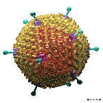
HIV and its life cycle

The saw no differences in virus diversity over 10 sequences per person, in 121 people,  71 of whom were in the placebo arm.  They did note, however, that linked transmissions showed less diversity in the env gene than normal – 1 vs 10%.  Over 75% of cases had a single founder virus, in both placebo and vaccine arms.  There was no significant divergence from the vaccine sequence in either group in anything but the Pro aa sequence – with some non-significant evidence for Env variation.

When they looked for Env sites under selection in gp120, they saw 4 in the placebo group at positions 181, 208, 327 and 359 – with less variation in vaccine than placebo recipients.  Rolland speculated that this could be to do with entry being more restrictive in vaccinees?  4 different sites in the vaccine group were under selection: they found that for MHC I epitopes there was a greater distance for vaccine than placebo groups, with a result that was not significant for MHC II epitopes.

There was a trend toward longer Env V2 loop sequences in vaccinees at later times, and a reduced number of cysteines in Env among vaccinees – this was seen also in the VAX004 trial.

Phil Berman – formerly of VaxGen, which made the gp120 for RV144 and earlier trials – mentioned that there was lower variance in Env than in the unsuccessful VAX 003 trial.  Jerome Kim noted that men seroconverting had a much higher incidence of HCV infection – which could be associated with undeclared IV drug use.

Katharine Barr of Univ Alabama spoke next, on the increased incidence of multiple variant transmission of HIV in VAX003 injection drug users.  She noted that this efficacy trial was of gp120 in IV drug users, while VAX004  was in MSM and high-risk women: they speculated that differences if any could be due to transmission route – as in, IV route vs sexual.  She further noted that in RV144, the best (non-significant) effect was in low-risk heterosexuals.

Something that was a little disturbing to me, given HIV transmission in our part of the world is overwhelmingly by heterosexual sex, was that the IV route is responsible for 10% of world infections.  They had looked at transmitted founder viruses – the ones going in and replicating in recipients.  They predicted that consensus of a low diversity lineage is the sequence of the founder virus – and that several founders would give multiple low variance lineages.

She noted that 80% of heterosexual infections are established by single viruses, so there exists a window of opportunity of viral vulnerability when vaccine-induced immunity could block infection.  However, with MSM, the multiple infection goes up to 40%; while injection drug users (IDUs) are less studied, multiplicity goes up  60% in one study and 31% in another….

Looking at Vax003 results, they had asked how high a barrier there had been for placebo infections, and whether in vaccinees there were more or fewer founder viruses?  While they had found that there was an 44% incidence of multiple variant transmission in the  placebo arm, and  22% in the vaccinees, this was unfortunately not significant, given the low numbers.  There was a median of 1.8 viruses per transmission vs 1.3, but this too was not significant.  However, it could mean there is a higher bar for vaccine protection among IDUs, which has important implications for which groups to use in vaccine trials.

Katherine incidentally gave the best answer yet heard to a long and detailed question: “I think that’s a really good question but I have zero data to address it…” = I don’t know.

Which prompted thoughts of new conference drinking games: take a shot every time you hear a speaker say “I would like to thank the organisers for inviting me…”, or “Our hypothesis [generally pronounced hy-PAH-the-sis] was…”, or a question which starts with either “…really good talk / great data” or “So – ummmm – when you/we did…”.

Paul Edlefsen (Fred Hutchinson Cancer Res Ctr) described a sieve analysis of RV144 [and started: “So…umm…” = another shot!].  He repeated the finding that observed correlates of risk generated two hypotheses; namely, that high IgG response to Env protected from HIV infection while a high IgA response interfered with protection.  Additionally, analysis of the antibody response using scaffold V region showed that a high V2 response correlated with a lower infection rate.  He noted that the STEP trial results showed a distinct difference in Gag between vaccine and placebo groups.  He noted further that were only 110 usable subjects in RV144, so they could only detect large sieve effects in their study of CTL and Ab epitope responses.

MUCH MIND-NUMBINGLY BORING STATISTICAL METHODOLOGY FOLLOWED…sorry, Paul!

There were 2 sites of evidence for sieving – aa positions 169 and 181 in the Env V2 loop, in the middle of a region identified by Ab binding array data.  There was also some evidence of covariation among pairs of aa residues in the V2 loop for vaccinees only.

After a long and complicated structural question, he gave the second-best answer of the conference: “I could say that I do, or that I don’t – but I have so little expertise in this area…(laughter)”.  And after long rambling statement: – “I’m sorry, was there a question in there?”

Brandon Keele (National Cancer Inst, MD) described work on NHPs which they had extended to studying human transmission of HIV, on transmitted/founder viruses.  NHP studies show multiple founders because doses are high generally, in order to get 100% infection rates.  One study using very low dose multiple intrarectal exposures to see if one can immunise macaques showed that one virus could do it.  Animals followed up from early times stayed with one evolving variant.

He noted that the consensus sequences in humans posited to have had one transmitted variant are average in  neutralisation susceptibility.  These viruses are all functional in vitro and in vivo and one can get full length viral clones ex NHPs which recap original founder viral load and pathogenicity.  All such viruses use the CCR5 coreceptor.  All HIV clones replicate in CD4 T-cells but not in  macrophages.  The transmission signature is to increase Env processing and infectivity.

They now mix cloned viruses with tags so can follow them in NHP challenge experiments, as most challenge studies have used virus with <1% diversity, which represents a clone in any one epitope – which he felt to be non-reflective of the real world .

The closing plenary session was opened by IAVI‘s Wayne Koff, who remarked that he had heard someone say “The  airport….”, in answer to the session name “Where do we go next?”….

Jeffrey Boyington (Vaccine Res Ctr) described some very impressive work on using structure of Env for rational immunogen design, specifically to target the CD4 binding site as a good target for broadly neutralising Ab.  They used crytallographic data to make proteins best mimicking the struc and then used them as immunogens.  They had used stabilised resurfaced gp120 with mutations around the binding site and isolated dozens of Abs with them from several infected subjects.  Part of the process involved stabilising flexible regions by bolstering cysteine content, removing glycans from the site of interest and adding them to immunodominant sites, and using Chikungunya virus VLPs to multimerise spike proteins for maximal immunogenicity.  Boyington noted that there were 80 native trimers on the surface of the VLPs, and that one can put the Outer Domain of gp120 on the tip of each monomer.  They get good Ab back for gp120 and get CD4 binding site Ab in rabbits.  In rhesus monkeys primed with gp140 trimers they got good boosting and better Abs to the CD4 BS.

Altogether a very impressive account – and one which advances to possibility of other opportunities for the design of other good broad-binding vaccine epitopes.

Rick King of IAVI followed, with an account of the current status and future directions of vector-based HIV vaccines.  He stated that most HIV vaccines now involve vectors – so there is a wealth of data that can be efficacious, so how to use it?  He thinks that we want the next generation of vectored vaccines to block infection and control virus load – meaning a combination of Ab and cellular responses.He noted that in NHPs, SIV protection is possible, and that it requires Env in the vaccine – and that the mechanism of protection is under intense investigation right now.

He further noted that in a DNA prime MVA boost vaccine regime, protection is associated with the avidity of the Abs.  Thus, a major goal is to improve the response to Env, by identifying the nature of the protective response, and enhancing and using native Envs to do it.  He stated in this context that there were only two vaccine regimens using native spike protein – and that one of them is the SA AIDS Vaccine Initiative (SAAVI) vaccine.

It was possible to engineer Env to bind a broader array of broadly neutralising Ab and to incorporate it into vesicular stomatitis virus (VSV) instead of the native G protein spike, or into canine distemper virus (CDV, a measles relative), which replicates in lymphoid tissue.  One could also bias processing of Env in CDV to get better cleavage and presentation.  The rCDV could be put into ferrets and shown to replicate.

He said that while the RV144 vaccine did not control viral load, vaccines can control SIV replication, so we need to have those components in HIV vaccines.  For instance, recombinant live cytomegalovirus (CMV) expressing the whole proteome of SIV could control the virus, this was associated with CD8 effector memory T-cells.

He thought we need to capitalise on information on mechanisms of control, and to increase immunity by use of replicating vectors and heterologous prime/boost combos, and deal with diversity by broadening the response.  The reason for replicating vectors was because live attenuated virus works for SIV – preventing infection and controlling replication.  Possibilities were vaccinia, measles, VSV, Sendai, CMV, AdV, CDV and VSV-HIV chimaeras.  As for diversity, one could increase the number of epitopes by using mosaics, and direct responses using conserved epitopes, as Tomas Hanke has demonstrated in IAVI-funded trials using chimpanzee Ad as prime then MVA as a boost with his HIVCONS Ag.

Finally, there was what I consider to have been the best talk of the conference – simply because it was much wider in scope than the rest: Steven Reed of the Infectious Disease Res Inst, Seattle, described new generation adjuvants for use with HIV.  He started by noting that adjuvants were necessary for lots of things; eg: for T-cell vaccines for TB and leishmania; for Ab response-broadening (Cervarix, HPV vaccine); Ag dose sparing (eg flu); to combat immune sensescence, and for vaccine therapy.

They had focused on a toll-like receptor (TLR4) agonist as an adjuvant, following work that showed that the well-known MPL was a TLR4 agonist ,and vaccines including TLR agonists had been used unknowingly since 1885.

He thinks the ideal adjuvant should have no effect on lymphocytes, no systemic effects, no non-specific B or T cell responses, should elicit potent long-lived responses, should redirect ongoing immune responses, and should be safe and effective in all age groups.  They had accordingly designed GLA – based on lipid A – to bind TLR4: this was purely synthetic, and induces Th1 CD4 helper cells and a broad humoral immunity.  They used a hexaacyl chain length that was preferred by human TLR4, which is restricted to macrophages and dendritic cells, has transient local effects, and reduces inflammation so as to get better central memory.

They can also formulate it differently for different vaccines and can get very different effects thereby.  For example, emulsion alone stimulates Th2 responses while GLA stimulates Th1 even in combo with an emulsion, which helps in leishmania and TB vaccines.

He noted that alum-based adjuvant stimulated mainly a Th2 response, while adding GLA gives a Th1 response with the same antigen.  They get good Ab diversity with GLA and expansion of it with the malaria vaccine – and Ab diversity leads to better neutralisation (eg transl med 2011).

GLA increases and broadens the haemagglutination-inhibtion (HAI) Ab response to the influenza vaccine Fluzone, which contains lots of inactivated virions.  He noted one gets a better protective response against “drifted” viruses – which have evolved away from the vaccine strains – with GLA.  Baculovirus-made H5N1 vaccine requires 30x less vaccine to get the same response with GLA.

It is also possible to get mucosal immunity by IM vaccination with HIV gp140, according to Robin Shattock’s results.

Reed noted that intradermal adjuvants are very rare – and that this looks good with flu vaccines delivered this way.  They were in the process of optimising the adjuvant formulation for intradermal delivery to increase vaccine potency, get mucosal immunity, and CD8+ T-cell responses.  Dermal dendritic cells have a wider range of TLRs than Langerhans cells – so Sanofi target them with ID delivery, and GLA works well to amplify the response.  It was impressive that they could protect ferrets with a single ID vaccine shot of flu vaccine.  It was also interesting that they are working with Medicago Inc., who have one of the most successful plant-produced influenza virus vaccine candidates, presently in human trial.

Thereafter, closing remarks from the conference organiser were as one would expect; people were honoured for their present and long-term contributions – notably Jose Esparza – and the venue of the next conference was announced to be Boston, with Dan Barouch as Organising Chair.

It was a good conference, with all of the high-intensity interactions and presentations one would expect from such a loaded topic.  However, it possibly suffered from over-emphasis of the “RV144 results” – which weren’t that impressive, in my opinion – as part of an effort to keep up perceived momentum from announcement of the RV144 success (small as it was) from the previous meeting.  For me, the highlights were the envelope antigen design talks, and what I managed to catch of the actual virology, and especially analysis of diversity by massively parallel sequencing.

We still don’t have an effective HIV vaccine – but we’re getting closer. 

From monkeys to humans, or…?

15 July, 2011

Adenovirus

A new Nature News item makes for interesting reading: it details how a new adenovirus, which had devastated a captive colony of titi monkeys, also jumped into a researcher working with them – and then into a family member who had had no other contact with the monkeys.

I put this comment up there:

While it may be unusual for adenoviruses to do this, there are a number of viruses which jump from monkeys to people – not the least of which are the HIVs.

This article, however, also raises to possibility that the virus may have gone the other way – that is, from humans to monkeys. This is also not that unusual; in fact, measles is a major risk factor in certain primate facilities, as certain monkeys can contract it easily, and often die.

The other possibility – that it came from a rodent or other animal – is potentially worrying, given that the virus was hitherto uncharacterised, and rodents tend to be ubiquitous.

Just goes to show: we really, really do need a Global Virome project, to pick up on all the little nasties out there.

MicrobiologyBytes Archive

14 December, 2007

Before I established this site, I posted a number of guest blogs to do with viruses on Alan Cann’s very wonderful MicrobiologyBytes site. Here are links to all the virus-related ones.

Maybe Not Quite The End

Posted on January 15, 2008
Review of a paper describing the receptor for the H5N1 HA protein

Given the current scare over H5N1 influenza virus in swans in the UK, it is possibly timely to recall that I wrote a little while ago in MicrobiologyBytes about how easy it appeared to be for […]

Bandicoot Blues

Posted on November 30, 2007
Description of a unique newly-described virus that looks like a chimaera of a papillomavirus and a polyomavirus

Now that the dust has begun to settle after the launch of Merck’s much-hyped Gardasil genital papillomavirus vaccine – discussed in MicrobiologyBytes here and here – people are turning again to looking at the natural history […]

Hurting rather than helping?

Posted on November 21, 2007
Some news on the failure of the Merck Adenovirus 5-vectored HIV vaccine

It should not have escaped the eye of the interested bystander that there has been a most unfortunate and premature end to a HIV vaccine trial recently – and that something that had been tested as […]

A Deeper Meaning

Posted on November 10, 2007
Some microbiology-related poetry….

I inadvertently became a published literary critic a little while ago. A long-time English Department colleague asked me for some help interpreting the collected works of possibly the most important modern poet from South Africa, and […]

Don’t look now, they’re in your genes

Posted on September 14, 2007
Description of natural insertions of virus gene fragments into a variety of organisms and how they elicit pathogen-derived resistance

And they’re protecting you! If you’re an insect, that is. Or possibly a plant.
In a remarkable convergence of news, an Israeli group led by Ilan Sela described how Israeli acute paralysis virus, which is implicated in […]

To bee or not to bee

Posted on September 11, 2007
News of how a single virus is suspected in the causation of “colony collapse disorder” of bee hives in the USA

A major recent mystery in US agriculture has been the phenomenon of “colony collapse disorder” (CCD) in honey bees. […]

This is the End

Posted on August 29, 2007
H5N1 highly pathogenic avian influenza virus mutates…

This is the End. Or the beginning of the end. Or possibly, the end of the beginning?
To misquote the immortal Bill Shankly: “It’s not a matter of life and death: it’s much more important than that”.
Having […]

Rolling down the road

Posted on August 27, 2007
Musings on rolling circle replication in viruses

In my idle moments (alas, too few these days!) I often try to think up lists of rock songs with a virus theme: you know, like “Cucumo” by the Beech Boys… “I got them ol’ burnin’, […]

Rooting the tree

Posted on August 3, 2007
News on inferring “ancestor sequences” for HIV to help make broadly effective vaccines

While fossilized viruses have never been found, we can often infer probable lines of evolutionary descent by analysis of extant genomic sequences. This sort of molecular phylogenetic approach has thrown up all sorts of interesting […]

It’s Life, Jim, but not as we know it…

Posted on July 24, 2007
Exploring what it means to be “alive”

Which could well apply to viruses, my very own favourite organisms – after all, they don’t respire, grow, excrete or any of those other good things […]

A feeling for the molechism*

Posted on June 26, 2007
Musings on what viruses are.

I think it’s permissible, after working on your favourite virus for over 20 years, to develop some sort of feeling for it: you know, the kind of insight that isn’t […]

Plus ça change, plus c’est … le same Web, only better?

Posted on June 8, 2007
A personal history of teaching Virology via the Web.

My, how things do change… I found myself reflecting, while I was looking over the detritus on our Web server of some 13 years of posting pages on the Web. “Orphan” pages, unconnected […]