Posts Tagged ‘virus’

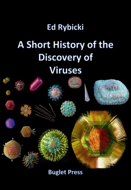
A Short History of the Discovery of Viruses

6 March, 2015

Now much updated, streamlined, added to and otherwise tarted up!  This is the Web version of an eBook, which you can now get here:

A Short History of the Discovery of Viruses – Edward Rybicki

Slide1

See also here for a “pocket reference version” of the discovery of viruses

Part 1: Filters and Discovery

Part 2: The Ultracentrifuge, Eggs and Flu

Part 3: Phages, Cell Culture and Polio

Part 4: RNA Genomes and Modern Virology

Sidebar 1: The Discovery of Filoviruses

Sidebar 2: Papillomaviruses and Human Cancer

Sidebar 3: Epstein-Barr Virus and Hepatitis B Virus

Sidebar 4: Human Retroviruses and Cancer

Sidebar 5: Maize Streak Virus: The Early History

Sidebar 6: Rinderpest and Its Eradication

Sidebar 7: Viruses and human cancer: the molecular age

Copyright Edward P Rybicki and Russell Kightley, February and March 2015, except where otherwise noted.

ViroBlogy: 2012 in review

1 February, 2013

So: thank you, anyone who clicked in, and regular visitors.  You make it worthwhile!!

The WordPress.com stats helper monkeys prepared a 2012 annual report for this blog.

Here’s an excerpt:

4,329 films were submitted to the 2012 Cannes Film Festival. This blog had 33,000 views in 2012. If each view were a film, this blog would power 8 Film Festivals

Click here to see the complete report.

Nonviral delivery of self-amplifying RNA vaccines – NOT!!

27 August, 2012

See on Scoop.itVirology and Bioinformatics from Virology.ca

“Despite more than two decades of research and development on nucleic acid vaccines, there is still no commercial product for human use. Taking advantage of the recent innovations in systemic delivery of short interfering RNA (siRNA) using lipid nanoparticles (LNPs), we developed a self-amplifying RNA vaccine. Here we show that nonviral delivery of a 9-kb self-amplifying RNA encapsulated within an LNP substantially increased immunogenicity compared with delivery of unformulated RNA. This unique vaccine technology was found to elicit broad, potent, and protective immune responses, that were comparable to a viral delivery technology, but without the inherent limitations of viral vectors. Given the many positive attributes of nucleic acid vaccines, our results suggest that a comprehensive evaluation of nonviral technologies to deliver self-amplifying RNA vaccines is warranted.”


I would re-ttitle this “Non-delivery of non-viral vectors…”.  Seriusly, folks, this is just the old Alphavax VEE vectors dressed up with with an in vitro synthesis step, and what amounts to a liposome delivery system.  Which means that it would be HIDEOUSLY expensive to produce, and is of no practical significance as a candidate vacine system whatsoever.


But I thank Alan Cann for pointing it out B-)

See on www.pnas.org

Recurvirostridae

23 August, 2012

See on Scoop.itVirology News

It’s a virus family!!  Or – a bird??  How confusing.  Someone needs to change their taxonomy.  Thanks to Russell Kightley for the keen observation.

See on en.wikipedia.org

Viruses linked to prostate cancer – ABC News (Australian Broadcasting Corporation)

1 August, 2012

See on Scoop.itVirology News

Australian scientists have made an important discovery about prostate cancer.

Nice little video: of course, they don’t say WHICH papillomaviruses are involved, along with Epstein-Barr virus.  Anyway – as long as it’s 16 and 18, good reason to get vaccinated, boys!  I thank Russell Kightley for sending me the clip.

See on www.abc.net.au

A feeling for the Molechism* – revisited

10 July, 2012

This is an update of a post I did on Alan Cann’s MicrobiologyBytes back in 2007, before i started ViroBlogy: I am doing this because (a) it’s mine, (b) I want to update it – and the MB version is archived, so I can’t.  So here we are again:

I think it’s permissible, after working on your favourite virus for over 20 years, to develop some sort of feeling for it: you know, the kind of insight that isn’t directly backed up by experiment, but that may very well be right. Or not – but in either case, it would take a deal of time and a fair bit of cash to prove or disprove, and would have sparked some useful discussion in the meantime. And then, of course, the insights you have into (insert favourite virus name here) – if correct – can usually be extended into the more general case, and if you are sufficiently distinguished, people may actually take them on board, and you will have contributed to Accepted Wisdom.

I can’t pretend – at least, outside of my office – to any such Barbara McClintock-like distinction; however, I have done a fair bit of musing on my little sphere of interest as it relates (or not) to the State of the Viral Universe, and I will share some of these rambles now with whomever is interested.

I have been in the same office now, and teaching the same course, more or less, for 32-odd years. In that time I have worked on the serology and epidemiology of the bromoviruses, cucumovirus detection, potyvirus phylogeny, geminivirus diversity and molecular biology, HIV and papillomavirus genetic diversity, and expressing various bits of viruses and other proteins in plants and in insect cells. However, much of my interest (if not my effort) in that time has been directed towards understanding how grass-infecting mastreviruses in particular interact with their environment and with each other, in the course of their natural transmission cycle.

Maize streak virus

Maxwell’s Demon (left, lower) and Martian Face (right, upper) visible on a MSV virion

Fascinating little things, mastreviruses: unique geminate capsid architecture, and at around a maximum of 2.8 kb of single-strand circular DNA, we thought they were the smallest DNA genomes known until the circoviruses and then the zoo of anello- and anello-like viruses were discovered. Their genomes code for only 4 proteins – two replication-associated, one movement and one capsid – yet we have managed to work on just one subgroup of mastrevirus species for 27 years, without exhausting its interest – at least, to us… (see PubMed list here). We also showed that one could see Martian faces quite distinctly on virions – and possibly even Maxwell’s Demon. But I digress….

Maize streak

Severe symptoms of MSV on sweetcorn

We have concentrated on the “African streak viruses” – related species Maize streak virus, Panicum streak virus, Digitaria streak virus, Sugarcane streak virus and friends – for two very simple reasons:
1. They occur in Africa, near us, and nowhere else;
2. Maize streak virus is the worst viral pathogen affecting maize in Africa.

So we get situational or niche advantage, and we get to work on an economically-important pathogen. One that was described – albeit as “…not of…contagious nature” – as early as 1901, no less.

Maize streak virus

Maize streak virus or MSV, like its relatives, is obligately transmitted by a leafhopper (generally Cicadulina mbila Naudé): this means we have a three-party interaction – of virus-host-vector – to consider when trying to understand the dynamics of its transmission. Actually, it’s more complicated than that: we have also increasingly to consider the human angle, given that the virus disease affects mainly the subsistence farming community in Africa, and that human activity has a large influence on the spread of the disease. So while considering just the virus – as complicated as that is – we have to remember that it is only part of the whole picture.

So how complicated is the virus? At first sight, not very: all isolates made from severe maize infections share around 97% of their genome sequence. However, however…that 3% of sequence variation hides a multitude of biological differences, and there is a range of relatives infecting grasses of all kinds, some of which differ by up to 35% in genome sequence. Moreover, maize is a crop plant first introduced to Africa a maximum of 500 years ago, so it is hardly a “natural” host – yet, all over Africa, it is infected by only a very narrow range of virus genotypes, from a background of very wide sequence diversity available.

So here’s an insight:

the host selects the virus that replicates best in it.

And lo, we found that in the Vaalharts irrigation area in the north of South Africa that the dominant virus genotype in winter wheat was a different strain – >10% sequence difference – to the one in the same field, in summer maize. Different grass species also have quite different strains or even species of streak viruses best adapted to them.

DendrogramNot all that profound a set of observations, perhaps, but they lead on to another insight:

streak viruses travel around as a cloud of variants or virus complex.

Not intuitively obvious, perhaps…but testable, and when we did, we found we were right: cloning virus genomes back out of maize or from a grass infected via leafhoppers gave a single predominant genotype in each case, with a number of other variants present as well. Looking further, we discovered that even quite different viruses could in fact trans-replicate each other: that is, the Rep/RepA complex of one virus could facilitate the replication of the genome of a virus differing by up to 35% in DNA sequence. We have also – we think – made nonsense of the old fancy that you could observe “host adaptation” of field isolates of MSV: we believe this was due to repeated selection by a single host genotype from the “cloud” of viruses transmitted during the natural infection cycle.

So, insight number three:

there is a survival benefit for the viruses in this strategy.

This is simple to understand, really, and relates to leafhopper biology as well as to host: the insects move around a lot, chasing juicy grasses, and it would be an obvious advantage to the streak virus complex to be able to replicate as a complex in each different host type – given that different virus genotypes have differential replication potential in the various backgrounds. This is quite significantly different, incidentally, to what happens with the very distantly-related (in terms of geological time) begomoviruses, or whitefly-transmitted geminiviruses: these typically do not trans-replicate each other across a gap of more than 10% of sequence difference.

Boring, I hear you say, but wait…. Add another factoid in, and profound insights start to emerge. In recent years, the cloud of protégés or virologist complex around me has accumulated to critical mass, and one of the most important things to emerge – apart from some frighteningly effective software for assessing recombination in viral genomes, which I wish he’d charge for – was Darren Martin’s finding that genome recombination is rife among African streak viruses. This was unexpected, given the expectation that DNA viruses simply don’t do that sort of thing; that promiscuous reassortment of components between genomes is a hallmark of RNA viruses. Makes sense in retrospect (an exact science), however, because of the constraints on DNA genomes: how else to explore sequence space, if the proof-reading is too good? And if you travel in a complex anyway…why not swap bits for biological advantage?

MSV web

Linkage map of the MSV genome, showing what interacts with what

So Darren swapped a whole lot of bits, in a tour-de-force of molecular virology, to create some 54 infectious chimaeric MSV genomes – and determined that

The pathogenicity of chimeras was strongly influenced by the relatedness of their parental viruses and evidence was found of nucleotide sequence-dependent interactions between both coding and intergenic regions“.

In other words –new insight:

the whole genome is a pathogenicity determinant, and bits of it interact with other bits in unexpected ways.

At this point you could say “Hey, all his insights are in fact hypotheses!” – and you would be partially correct, except for

Profound Insight No. 1hypotheses are the refuge of the linear-thinking.

Or its variant, found on my office wall:

“**c* the hypotheses, let’s just discover something”. I also have

“If at first you don’t succeed, destroy all evidence that you tried” and a number of exotic beer bottle labels on my wall – but I digress….

As an aside here, I am quite serious in disliking hypothesis-driven science: I think it is a irredeemably reductionist approach, which does not easily allow for Big Picture overviews, and which closes out many promising avenues of investigation or even of thought. And I teach people how to formulate them so they can get grants and publications in later life, but I still think HDS is a tyranny that should be actively subverted wherever possible.

Be all this as it may, now follows

Profound Insight No. 2genome components may still be individually mobile even when covalently linked.

Now take a moment to think on this: recombination allows genes to swap around inside genetic backgrounds so as to constitute novel entities – and the “evolutionary value of exchanging a genome fragment is constrained by the number of ways in which the fragment interacts with the rest of the genome*“. Whether or not the genome is RNA, DNA, in one piece or divided. All of a sudden, the concept of a “virus genome” as a gene pool rather than a unitary thing becomes obvious – and so does the reductionism inherent in saying “this single DNA/RNA sequence is a virus”.

So try this on for size for a brand-new working definition of a virus – and

Profound Insight No. 3a virus is an infectious acellular entity composed of compatible genomic components derived from a pool of genetic elements.

Sufficiently paradigm-shifting for you? Compare it to more classical definitions – yes, including one by AJ Cann, Esq. – and see how much simpler it is. It also opens up the possibility that ANY virus as currently recognised is simply an operational assembly of components, and not necessarily the final article at all.

Again, my favourite organisms supply good object examples: the begomoviruses – whitefly-transmitted geminiviruses –

  • may have one- or two-component genomes;
  • some of the singleton A-type components may pick up a B-type in certain circumstances;
  • some doubletons may lose their B without apparent effect in model hosts;
  • some A components may apparently share B components in natural infections;
  • the A and B components recombine like rabbits with cognate molecules (or Bs can pick up the intergenic region from As);
  • in many cases have one or more satellite ssDNAs (β DNA, or nanovirus-related components) associated with disease causation;

…and so on, and on…. An important thing to note here is the lab-rat viruses – those isolated early on, and kept in model plant species in greenhouses – often don’t exhibit any of these strangenesses, whereas field-isolated viruses often do.

Which tells you quite a lot about model systems, doesn’t it?

But this is not only true of plant viruses: the zoo of ssDNA anello-like viruses found in humans and in animals – with several very distantly-related viruses to be found in any individual, and up to 80% of humans infected – just keeps on getting bigger and weirder. Added to the original TT virus – named originally for the initials of the Japanese patient from whom it was isolated, and in a post hoc exercise of convoluted logic, named Torque teno virus (TTV) [why don’t people who work with human or animal viruses obey ICTV rules??] – are now Torque teno minivirus (TTMV) and “small anellovirus” SAV) – all of which have generic status. And all of which may be the same thing – as in, TTVs at a genome size of 3.6–3.8 kb may give rise to TTMVs (2.8-29 kb) and SAVs (2.4-2.6 kb) as deletion mutants as part of a population cloud, where the smaller variants are trans-replicated by the larger. Thus, a whole lot of what are being described as viruses – without fulfilling Koch’s Postulates, I might point out – are probably only “hopeful monsters” existing only as part of a population. Funnily enough, this sort of thing is much better explored in the ssDNA plant virus community, given that working with plant hosts is so much easier than with human or animal.

And now we can go really wide, and attempt to be profound on a global scale: it should not have escaped your notice that the greatest degree of diversity among organisms on this planet is that of viruses, and viruses that are found in seawater in particular. There is a truly mind-boggling number of different viruses in just one ml of seawater taken from anywhere on Earth, which leads respectable authors such as Curtis Suttle to speculate that viruses almost certainly have a significant influence on not only populations of all other marine organisms, but even on the carbon balance of the world’s oceans – and therefore of the planet itself.

Which leads to the final, and most obvious,

Profound Insight (No. 4)in order to understand viruses, we should all be working on seawater…. 

That is where the diversity is, after all; that is where the gene pool that gave rise to all viruses came from originally – and who knows what else is being

Hypolith – cyanobacteria-derived, probably – under a piece of Namib quartzite from near Gobabeb Research Station

cooked up down there?

And this is the major update: not only have I managed to get funded for a project on “Marine Viromics” from our local National Research Foundation – a process akin to winning the lottery, and about as likely to succeed – I am also collaborating with friends and colleagues from the Institute for Microbial Biotechnology and Metagenomics at the University of the Western Cape on viruses in desert soils, and associated with hypoliths– or algal growths found under quartzite rocks in extreme environments.

Thus, I shall soon be frantically learning how to deal with colossal amounts of sequence data, and worse, learning how to make sense of it.  We should have fun!

——————————————————————————————————————–

* And as a final curiosity, I find that while I – in common with the World Book Encyclop[a]edia and Learning Resources – take:mol|e|chism or mol|e|cism «MOL uh KIHZ uhm», noun. to mean any virus, viewed as an infective agent possessing the characteristics of both a living microorganism and a nonliving molecule; organule.
[molechism < mole(cule) + ch(emical) + (organ)ism; molecism < molec(ule) + (organ)ism] –
There is another meaning… something to do with sacrifice of children and burning in hellfire eternally. This is just to reassure you that this is not that.

Science| Special Issue: H5N1 [exploring the “supervirus” controversy]

29 June, 2012

See on Scoop.itVirology News

“Introduction
The publication in this issue of these research papers on the airborne tranimssion [sic] of H5N1 marks the end of 8 months of controversy over whether some of the data, now freely accessible, should be withheld in the public interest.”


I think this is an important landmark in the so-called “dual use” debate: that is, the propensity of bodies in the US to attempt to regulate the release of information that MAY be usable in the making of bioweapons, or be usable in bioterror attacks.


Let us diffidently point out at this juncture that it is only really the superpowers who are definitively known in recent years to have had bioweapons programmes – apart from apartheid-era South Africa, that is! – and that damn nearly ANYTHING published on transmission or mechanisms of pathogenicity of human or animal pathogens (or even plant, for that matter) could be termed “dual use” if someone wanted to – and censored as a result.


It is also – as I tire of pointing out – possible to PROTECT against H5NX viruses using conventional vaccines right now – and the new universal flu vaccines coming on stream will almost certainly make this even more feasible.


The fact is that H5N1 flu is an ever-present threat to people living in Egypt, Indonesia, Cambodia, Viet Nam, Thailand and China – WITHOUT being weaponised.  It is no more than a notional threat to the US or Europe – and keeping information that could help in understanding how or how soon the virus could mutate to pandemicity out of people’s hands, is simply stupid. 

See on www.sciencemag.org

Endogenous RNA viruses of plants in insect genomes

5 June, 2012

See on Scoop.itVirology News

“Endogenous viral elements (EVEs) derived from RNA viruses with no DNA stage are rare, especially those where the parental viruses possess single-strand positive-sense (ssRNA +) genomes. Here we provide evidence that EVEs that share a sequence similarity to ssRNA + viruses of plants are integrated into the genomes of a number of insects, including mosquito, fruit flies, bees, ant, silkworm, pea aphid, Monarch butterfly, and wasps. A preliminary phylogenetic analysis places these EVEs as divergent relatives of the Virgaviridae and three currently unclassified plant viral species.”

I have covered this before, in ViroBlogy, (and here, in 2007)as an interesting and probably under-appreciated phenomenon.  I note Eddie Holmes and colleagues have now taken it much, much further – which incidentally lends significant credence to my supposition that virus/vector/plant coevolution was probably a fair bit more intimate than has been supposed, with the newly-emerged (in evolutionary terms) insects and their viruses meeting terrestrial plants and THEIR viruses.  And mixing everything up, as I have speculated elsewhere (Origins of Viruses).

I thank Jean-Marie Verchot for drawing my attention to this!

See on www.sciencedirect.com

Silencing is golden!

18 April, 2012

An excellent journal club article by Mark Whitehead:

Pavan Kumar, Sagar Subhash Pandit, Ian T. Baldwin. Tobacco Rattle Virus Vector: A Rapid and Transient Means of Silencing Manduca sexta Genes by Plant Mediated RNA Interference. PLoS ONE, 2012; 7 (2): e31347 DOI: 10.1371/journal.pone.0031347

Specific Insect Gene Silencing achieved by ingestion of plant produced dsRNA, via a transient viral vector platform.

RNAi- mediated gene silencing is an endogenous mechanism and has been utilised in reverse genetics in a number of organisms and it has the potential to be used as a tool for pest control.

The diagram below gives a good summary on the standard RNAi process. Briefly,  dsRNA produced in the nucleus is transported to the cytoplasm; alternatively, exogenous dsRNA can be taken up by cells with the help of a cell surface protein. In the cytoplasm, dsRNA is cleaved by RNaseIII type enzymes (dicers) to produce approximately 22 bp fragments, called small interfering RNAs (siRNAs). One strand of the siRNA (guide strand) is incorporated into the RNA-induced silencing complex (RISC) with the perfectly complementary site in a target mRNA to form a guide strand-target mRNA duplex. The target mRNA is then sliced by the Argonaute protein of RISC.

(With permission from http://www.RNAiweb.com.)

Plants have RNA-dependant RNA polymerases (RdRPs) that accentuate the process as they extend the bound guide strand to create more dsRNA that can then re-enter the RNAi cycle. dsRNA delivered to insects by various routes has been seen to induce RNAi, however insects lack RdRPs and therefore require a large constant supply of siRNAs for sustained gene silencing. Herbivorous insects feeding on stably transformed transgenic host plants have been seen to take up the produced dsRNA molecules into their gut cells, causing post transcriptional gene silencing. Generation of these stable transgenic plant lines is a time consuming task, while transient plant transformation offers a faster and more versatile approach, allowing for a number of dsRNA products to be created as a quicker screening method.

Larvae of the tobacco hornworm Manduca sexta contain genes that encode for nicotine-catabolising enzymes, rendering them resistant to the toxic nicotine alkaloid produced by their host plant Nicotiana attenuata. It was previously seen that some cytochrome P450 (CYP) genes were up-regulated in the larval gut in response to nicotine ingestion (CYP4M1 and CYP4M3 genes) and CYP6B46 was down-regulated when fed on nicotine suppressed plants.

In this paper the tobacco rattle virus (TRV) was used to transiently produce dsRNAs in Nicotiana attenuata – this approach was termed plant-virus based dsRNA producing system (VDPS) – in comparison to stably transformed plants – termed plant mediated RNAi (PMRi) for the silencing of these lepidopteran genes.

They initially checked to see if M. sexta could indeed take up the dsRNA and cause PMRi. It was observed that when the larvae were fed on a transgenic plant expressing dsRNA for the CYP6B46 gene, there was CYP6B46 smRNA found in the midgut and a reduction in the CYP6B46 transcript levels was observed, effectively causing silencing of the gene. It was very specific as the transcript levels of a similar gene (CYP6B45 – 80% similarity) was not affected. The VDPS was tested and compared to the PMRi for the same target and produced comparable silencing, that was also highly specific and did not cause any “off-target” effects.

Since the VDPS is a more rapid technique and was seen to be comparable to the PMRi, it was therefore used to screen the other gene targets – CYP4M1 and CYP4M3. Again the smRNAs for each were seen to be present in the midgut of the larvae when fed on the plants and the transcript levels were reduced with high specificity. The reduction of the CYP4M3 transcription levels also caused larval growth to decrease, indicating that this gene may a central role in nicotine tolerance.

The length of the dsRNA is known to have an effect on RNAi experiments and it would be ideal if the lengths were standardised. It is possible that the lepidopteran dicers that function in extremely alkaline environments of the midgut are specialized and possess different dicing properties than the plant dicers; consequently, insect-dicer diced smRNA might be more effective than the plant-dicer diced smRNA in gene silencing in insects.

Plant Dicers (DCLs) are involved in the biogenesis of smRNA by cleaving longer dsRNA. Four different types of DCLs are reported in higher plants. Their function has been found to overlap in plants, suggesting that one DCL can contribute to and/or compensate for the function of the others. Hence, more than one DCL might be involved in processing long dsRNA.

To address this they then silenced different combinations of the four N. attenuata’s Dicer genes in the transgenic PMRi lines producing CYP6B46 dsRNA. Long CYP6B46 transcript levels in the plants was found to be increased more than 50 fold when the DCL 1,3,4 or DCL 2,3,4 were co-silenced. These then lead to an enhanced silencing effect in the larvae midgut, indicating that there could be a preference for insect diced smRNAs or simply that the larger dsRNAs were more stable and the higher concentration enhanced the silencing effect. It also suggests that the plant and insect RNAi machinery respond differently to the dsRNA.

In conclusion PMRi can be a specific and robust system of gene silencing in M. sexta. PMRi would be the method of choice for crop protection in countries which allow the growth of transgenic crops. While retaining all the virtues of PMRi, VDPS promises to be a rapid and high throughput alternative, suitable for ecological research.

This article has been a short review of the journal article stated below. For more in depth information on this research, follow the link and download the freely available journal article.

A Short History of the Discovery of Viruses – Part 1

6 February, 2012

The following text has now appeared in modified form in an ebook, for sale for US$4.99 on the iBooks Store

A Short History of the Discovery of Viruses

While people were aware of diseases of both humans and animals now known to be caused by viruses many hundreds of years ago, the concept of a virus as a distinct entity dates back only to the very late 1800s.  Although the term had been used for many years previously to describe disease agents, the word “virus” comes from a Latin word simply meaning “slimy fluid”.

Porcelain filters and the discovery of viruses

The invention that allowed viruses to be discovered at all was the Chamberland-Pasteur filter.  This was developed in 1884 in Paris by Charles Chamberland, who worked with Louis Pasteur.  It consisted of unglazed porcelain “candles”, with pore sizes of 0.1 – 1 micron (100 – 1000 nm), which could be used to completely remove all bacteria or other cells known at the time from a liquid suspension.  Though this simple invention essentially enabled the establishment of a whole new science – virology – the continued development of the discipline required a string of technical developments, which I will highlight as appropriate.

Pasteur Germ Proof Filter, c. 1890, Pasteur-Chamberland Filter Co., Dayton, Ohio – Museum of Science and Industry (Chicago)


As the first in what was to be an interesting succession of events, Adolf Eduard Mayer from Germany, publishing in 1886 on work done in Holland from 1879, showed that the “mosaic disease” of tobacco – or “mozaïkziekte”, as he named it in his paper – could be transmitted to other plants by rubbing a liquid extract, filtered through paper, from an infected plant onto the leaves of a healthy plant.  However, he came to the erroneous conclusion that it must be a bacterial disease.

The first use of porcelain filters to characterize what we now know to be a virus was reported by Dmitri Ivanovski in St Petersburg in Russia, in 1892.  He had used a filter candle on an infectious extract of tobacco plants with mosaic disease, and shown that it remained infectious: however, he concluded the agent was probably a toxin as it appeared to be soluble.

The Dutch scientist Martinus Beijerinck in 1898 reported similar experiments with bacteria-free filtered extracts, but made the conceptual leap and described the agent of mosaic disease of tobacco as a “contagium vivum fluidum”, or contagious living fluid, because he was convinced the infectious agent had a liquid nature. The extract was completely sterile, could be kept for years, but remained infectious.  The term virus was later used to describe such fluids, also called “filterable agents”, which were thought to contain no particles.  The virus causing mosaic disease is now known as Tobacco mosaic virus (TMV).  A paper commemorating Ivanovsky and Beijerinck’s work – “One Hundred Years of Virology” – was published in Journal of Virology 1992 to honour both pioneers.

The first animal viruses

The second virus discovered was what is now known as Foot and mouth disease virus (FMDV) of farm and other animals, in 1898 by the German scientists Friedrich Loeffler and Paul Frosch.  Again, their “sterile” filtered liquid proved infectious in calves, providing the first proof of viruses infecting animals – a fact commemorated by an article in 1998 in the Journal of General Virology.  Indeed, some believe that the true discoverers of viruses were these two scientists, as they concluded the infectious agent was a tiny particle, and was not a liquid agent.  The two went further by showing that it was possible to vaccinate cows and sheep against the disease using filtered vesicle extract that had been heated sufficiently to destroy its infectivity: this was possibly the first use of an inactivated virus as a prophylactic vaccine.

In 1898 G Sanarelli, working in Uruguay, described the smallpox virus relative and tumour-causing myxoma virus of rabbits as a virus – but on the basis of sterilisation by centrifugation rather than by filtration.

The first human virus: yellow fever

The first human virus described was the agent which causes yellow fever: this probably originated in Africa, but was spread along with its mosquito vector Aedes aegyptii to the Americas and neighbouring islands by the slave trade.  Indeed, the  declaration of independence from France by Haiti in 1804 was made possible in part by the devastating effect of the disease on the French army sent to quell a slave revolt there. The virus was discovered and reported in 1901 by the US Army physician Walter Reed, after pioneering work in Cuba by Carlos Finlay reported in 1881 hypothesising that mosquitoes transmitted the deadly disease

The agent became the subject of intense study because, in the Spanish-American war in Cuba in the 1890s, about 13 times as many soldiers died of yellow fever as died from wounds. The subsequent eradication of mosquitoes in Panama is probably what allowed the completion of the Panama Canal – stalled because of the death rate among workers from yellow fever and malaria.

Rinderpest and rabies

The paper describing rinderpest as a virus disease

A finding that was later to have great importance in veterinary virology was the discovery by Maurice Nicolle and Adil Mustafa (also known as Adil-Bey), in Turkey in 1902, that rinderpest or cattle plague was caused by a virus.  This had been for several centuries the worst animal disease known worldwide in terms of mortality: for example, an epizootic or animal epidemic in Africa in the 1890s that had started in what is now Ethiopia in 1887 from cattle imported from Asia, had spread throughout the continent by 1897, and killed 80-90% of the cattle and a large proportion of susceptible wild animals in southern Africa.  Many thousands of people died of starvation as a result.  The virus is, incidentally, only the second to have ever been eradicatednearly 100 years after its discovery.

Viruses and Vaccines

Sir Arnold Theiler, a Swiss-born veterinarian working in South Africa, had been appointed as state veterinarian for the Zuid-Afrikaansche Republiek prior to 1899, on the strength of his having produced a smallpox vaccine for miners in the Johannesburg area.  He then developed a crude vaccine against rinderpest by 1897, without knowledge of the nature of the agent: this consisted of blood from an infected animal, injected with serum from one that had recovered – something also shown to work with FMDV by Loeffler and Frosch.  This risky mixture worked well enough, however, to eradicate the disease in the region.  He went on to do the same thing successfully for African horsesickness virus and other disease agents, in an institute (Onderstepoort Veterinary Institute) that still works on the virus.

The description in Annales de l’Institut Pasteur by Remlinger and Riffat-Bay in 1903 of the agent of rabies as a “filterable virus was the culmination of many years of distinguished work in France on the virus, started by Louis Pasteur himself. While Remlinger credited Pasteur with having the notion in 1881 that rabies virus was an ultramicroscopic particle, the fact is that Pasteur and Emile Roux had also, in 1885, effectively made a vaccine against rabies by use of dried infected rabbit spinal cords, without any knowledge of what the agent was.

Title page of the original article in Annales de l’Institut Pasteur Volume 17 of 1903

It is interesting that the same volume of the Annales which reported the rabies agent also has a discussion on whether or not the smallpox agent variola virus and the vaccine against it, vaccinia virus, were differently-adapted variants of the same thing, or were different viruses.

More and more viruses

The viral nature of many disease agents started to be made evident around this time, as more researchers started investigating known diseases.  In 1904, E Baur in Germany described an infectious variegation of Abutilon that could only be transmitted by grafting, that was not associated with visible bacteria.  This is now known to be due to Abutilon mosaic virus, now known to be a single-stranded DNA geminivirus.

Abutilon mosaic

Incidentally, the earliest recorded description of a plant disease was probably in a poem in 752 CE by the Japanese Empress Koken, describing symptoms in eupatorium plants.  It was shown in 2003 that the striking yellow-vein symptoms were caused by a geminivirus infection.

Interestingly, also in 1906, A Zimmermann proposed – in a paper entitled “Die Krauselkrankheit des Maniok” – that the agent of mosaic disease of cassava that had first been described from German East Africa (now Tanzania) in 1894, was a filterable virus.  This was the second geminivirus discovered, although this was only proved in the 1970s.

Cassava affected by a recombinant African cassava mosaic virus in western Kenya, 1997

Cassava affected by a recombinant African cassava mosaic virus in western Kenya, 1997.  Insets, from left: healthy cassava, mild disease, severe disease

In 1906, Adelchi Negri – who had previously discovered the Negri bodies in cells infected with rabies virus – showed that vaccinia virus, the vaccine for the dreaded smallpox caused by variola virus, was filterable.  This was the final step in a long series of discoveries around smallpox, that started with Edward Jenner’s use of what was supposedly cowpox, but may have been horsepox virus to protect people from the disease in 1796.

An Egyptian stele thought to represent a polio victim (1403–1365 BC). Note the characteristic withering of one leg.

An Egyptian stele thought to represent a polio victim (1403–1365 BC). Note the characteristic withering of one leg.

The disease now known as poliomyelitis was first clinically described in England in 1789, as “a debility of the lower extremities”.  However, it had been known since ancient times, and had even been depicted clearly in an Egyptian painting from over 3000 years ago.

An important development in human virology in 1908, therefore, was the finding by Karl Landsteiner and Erwin Popper in Germany that poliomyelitis or infantile paralysis in humans as it was known then, was caused by a virus: they proved this by injecting a cell-free extract of a suspension of spinal cord from a child who had died of the disease, into monkeys, and showing that they developed symptoms of the disease.

Viruses and cancer

In 1908, Oluf Bang and Vilhelm Ellerman in Denmark were the first to associate a virus with leukaemia: they successfully used a cell-free filtrate from chickens with avian leukosis to transmit the disease to healthy chickens.

The first solid tumour-causing virus, or virus associated with cancer, was found by Peyton Rous in the USA in 1911.  He showed that chicken sarcomas, or solid connective tissue tumours, could be transmitted by grafting, but also that a filterable or cell-free agent extracted from a sarcoma was infectious.  The virus was named for him as Rous sarcoma virus, and is now known to be a “retrovirus”,as is chicken leukaemia virus,in the same virus family as HIV.

Eaters of Bacteria: The Phages

"Twort" by Obituary Notices of Fellows of the Royal Society, Vol. 7, No. 20. (Nov., 1951), pp. 504-517.. Licensed under Public Domain via Wikimedia Commons - http://commons.wikimedia.org/wiki/File:Twort.jpg#mediaviewer/File:Twort.jpg

“Twort” by Obituary Notices of Fellows of the Royal Society, Vol. 7, No. 20. (Nov., 1951), pp. 504-517.. Licensed under Public Domain via Wikimedia Commons – http://commons.wikimedia.org/wiki/File:Twort.jpg#mediaviewer/File:Twort.jpg

Two independent investigations led to the important discovery of viruses that infect bacteria. In 1915, Frederick Twort in the UK accidentally found a filterable agent that caused the bacteria he was growing to lyse, or burst open.  Although he showed that it could pass through porcelain filters, and could be transmitted to other colonies of the same bacteria, he was not sure whether or not it was a virus, and referred to it as “the bacteriolytic agent”.  It is interesting that he was actually attempting to grow vaccinia virus in culture, and that it was a contaminating staphylococcus that he noticed was being lysed by his infectious agent.

www_sciencedirect_com_science__ob_PdfExcerptURL__imagekey_1-s2_0-S0140673601203833-main_pdf__piikey_S0140673601203833__cdi_271074__orig_article__zone_centerpane__fmt_abst__eid_1-s2_0-S0140673601203833__user_635696_md5_610868932f41b70483e225

The original article published by Twort in The Lancet in 1915

Subsequently, Félix d’Hérelle in Paris published in 1917 that he had discovered a virus that lysed a bacterial agent he was culturing in liquid broth – a Shigella – that caused human dysentery, or diarrhoea.  He named the virus “bacteriophage”, or eater of bacteria, derived from the Greek term “phagein”, meaning to eat.  He showed a number of interesting properties of his shigella-specific bacteriophages, including that they could be adapted to other Shigella species or types by passaging them repeatedly, and that they protected rabbits against infection by lethal doses of bacteria

D’Hérelle’s main interest in his new discovery was in using them as a therapeutic agent for bacterial infections in humans: sadly, this idea did not take off in Europe or the Americas, largely due to the unreliability of the ill-understood phage preparations, although it was extensively exploited in the former USSR. Indeed, he mentored George Eliava who went on to found the Eliava Institute in Tbilisi, Georgia, which became a major centre for the use of bacteriophage cocktails against persistent bacterial infections in humans.  A review on phage therapy from the Institute was recently published to mark the centenary of Twort’s discovery in 1915. An excellent – if slightly childish – animation describing phages and phage therapy can be seen here.

D‘Hérelle’s main interest in his new discovery was in using them as a therapeutic agent for bacterial infections in humans: sadly, this idea did not take off in Europe or the Americas, although it was extensively exploited in the former USSR.

The 1896 paper from Annales de l’Institut Pasteur

Interestingly, and as reported in ViroBlogy previously, what could have been the first discovery of phages was probably described by Ernest Hankin, who had previously proved in India that cholera was caused by bacteria.  In 1896 in Annales de l’Institut Pasteur, he documented that river water downstream of cholera-infested towns on the Jumma river in India contained no viable Cholera vibrio – and that this was a reliable property of the water, and was probably responsible for limiting the spread of cholera

While he did not prove the presence of a “filterable agent”, he was recognised by d’Hérelle and others as having contributed to the discovery of bacteriophages.  In fact, d’Hérelle went to India in 1927, and put cholera phage preparations into wells in villages with cholera patients: apparently the death toll went down from 60% to 8%.

Influenza A viruses in waterbirds – Russell Kightley Media

The virus as human plague: the Spanish Flu

Possibly the worst human plague the world has ever seen swept across the planet between 1918 and 1922: this was known as the Spanish Flu, from where it was first properly reported, and it went on to kill more than 50 million people all over the world.  We now know it to have been H1N1 influenza type A: modern reconstruction of the virus from archived tissue samples and frozen bodies found in permafrost has shown it probably jumped directly into humans from birds.

Most medical authorities at the time thought the disease was caused by bacteria – however, MJ Dujarric de la Rivière, and Charles Nicolle – brother of Maurice – and Charles  Lebailly in France, separately proposed in 1918 that the causative agent was a virus, based on properties of infectious extracts from diseased patients.  Specifically, they found that the infectious agent derived from bronchial expectoration of an infected person was filterable, caused disease in monkeys via nasal administration and human volunteers via subcutaneous injection, and was not present in the blood of an infected monkey.  However, many scientists at the time still doubted that influenza was a viral disease – despite this contemporary comment in the British Medical Journal of 1918.

Conclusions from the Nicolle and Lebailly paper

Translation of this passage (courtesy of Mrs Francoise Williamson):

“Conclusions.

1⁰ The bronchial  expectoration of people suffering from flu, collected during the acute period, is virulent.
2⁰   The monkey (M. cynomolgus)  is sensitive to the virus  by sub-conjunctival and nasal inoculation.
3⁰   The flu agent is a filterable organism.  The inoculation  of the filtrate has indeed reproduced the illness in two of the people injected subcutaneously;  on the other hand when given intravenously it  appears to be ineffective. (two failures out of two tries).
4⁰ It is possible that the influenza virus does not occur in the patient’s blood.  The blood of a monkey with influenza, inoculated subcutaneously, did not infect man;  the negative blood result of subject 2 at D, is however, not convincing, the blood route seeming to be ineffective for  the flu virus transmission.”

Other agents of other diseases were found to be “filterable viruses” in the 1920s, including yellow fever virus by Adrian Stokes in 1927, in Ghana.  Indeed, the US bacteriologist and virologist Thomas Rivers in 1926 counted some sixty-five disease agents that had been identified as viruses.

Virus Assays: Counting the Viruses in the 1920s

The discovery of bacteriophages was a landmark in the history of virology, as it meant that for the first time it was relatively easy to work with viruses: many kinds of bacteria could be grown in solid or liquid culture quite easily, and the life cycle of the viruses could be studied in detail.  In fact, this later led to the birth of molecular biology, as described here

However, the beauty of working with phages was that they could be assayed – or counted in terms of infectious units – so easily, either by the plaque technique or by infections of liquid cultures.  This was not true of viruses of plants or of animals in the absence of similar culture techniques; these could only be assayed in a much more crude method using whole organisms.  One such method was by determining infection endpoints by serial dilution of inoculum, such as the now-famous ID50, or dose infecting 50% of the experimental subjects.

This changed in 1929 for plant viruses, with the demonstration by the plant virus pioneer FO Holmes that local lesions caused by infection of particular types of tobacco by TMV could be used as a means of assaying the infectivity of virus stocks.  This was then extended to other virus/host combinations, and allowed the rapid and quantitative assay of virus stocks – which, as it had done for phages, allowed the study of the properties of plant viruses, and led to their biological isolation and then purification.

TMV-induced local lesions in N. tabacum cv. glutinosa

TMV-induced local lesions in N. tabacum cv. glutinosa

Eggs and animals for virus culture

Chicken eggs for virus growth and assay

Possibly the next most important methodological development in virology after the discovery of phages was the proof that embryonated or fertilized hen’s eggs could be used to culture a variety of important animal and human viruses.  Ernest Goodpasture, working at Vanderbilt University in the USA, showed in 1931 that it was possible to grow fowlpox virus – a relative of smallpox – by inoculating the chorioallantoic membrane of eggs, and incubating them further. 

egg

While tissue culture had in fact been practiced for some time – for example, as early as the 1900s, investigators had grown “vaccine virus” or the smallpox vaccine now called vaccinia virus in minced up chicken embryos suspended in chicken serum – this technique represented a far cheaper and much more “scalable” technique for growing pox- and other suitable viruses.

An important first in a chain of related discoveries was the one by Howard Andervost, at Harvard University in 1929, who showed that human herpes simplex virus could be cultured by injection into the brains of live mice.

This led to the demonstration in 1930 by the South African-born Max Theiler – son of Sir Arnold – also at Harvard, that yellow fever virus could be similarly cultured: this allowed much easier handling of the virus, which until then had to be injected into monkeys in order to multiply it in their livers.  In addition, it allowed the development of attenuated or weakened strains of virus, by him and in parallel by a French laboratory, by serial passage or repeated transmission of the virus between mice. He also incidentally caught yellow fever from one of his mice through a laboratory accident. Culturing in mouse brains also allowed the successful animal testing of vaccine candidates, and of protective antisera, for which Theiler was awarded the Nobel Prize in 1951.  

Until 2008, this was the first and only recognition of virus vaccine work by the Nobel Foundation.

A consequence of this work was the landmark in medical virology that was the development of human vaccines against yellow fever virus, by Wilbur Sawyer in the USA in 1931: this followed on Theiler’s mouse work in using brain-cultured virus plus human immune serum from recovered patients to immunize humans – very similar to Theiler Senior’s strategy with rinderpest, more than thirty years later.

here for Part 2: The Ultracentrifuge, Eggs and Flu

here for Part 3: Phages, Cell Culture and Polio

and here for Part 4: RNA Genomes and Modern Virology

Copyright Edward P Rybicki and Russell Kightley, February 2012, except where otherwise noted.