Archive for March, 2015

Rinderpest and its eradication

30 March, 2015

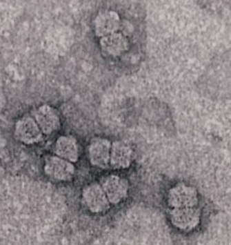
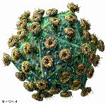
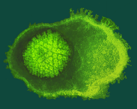
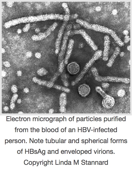
Rinderpest virus infects cattle, buffalo and several species of antelope among other animals: it is a member of the genus Morbillivirus, family Paramyxoviridae, and is related to measles and mumps viruses in humans, distemper virus in dogs, and a variety of relatively newly-described viruses in marine mammals.  It also almost certainly gave rise to measles virus sometime around the 11th-12th centuries CE, as an originally zoonotic infection – sourced in domestic animals – took root in humans and began to be passed around (see here).

Wikipedia describes rinderpest virus as

“…an infectious viral disease of cattle, domestic buffalo, and some species of wildlife. The disease was characterized by fever, oral erosions, diarrhea, lymphoid necrosis, and high mortality.”  

And:

“The term Rinderpest is taken from German, and means cattle-plague.”

“Cattle plagues” have occurred throughout recorded history, often associated with the large-scale movement of animals accompanying military campaigns.  Europe was badly hit in the 1700s, with three epizootics in 1709–1720, 1742–1760, and 1768–1786, and a major British outbreak in 1865/66.

Of particular interest in South African folklore, an outbreak in the 1890s killed up to 90 percent of all cattle in southern and north-west Africa, and resulted in the deaths of many thousands of people who depended on them. It was devastating enough that people still remember it as a legendary time of hardship. Sir Arnold Theiler was instrumental in developing a vaccine that curbed the epidemic – by simultaneously injecting animals with blood from an infected animal and antiserum from a recovered animal. This protected animals for long enough to allow their immune systems to respond to the virus, but was rather risky, even though it was used for several decades.

In the 1920s J. T. Edwards in what is now the Indian Veterinary Research Institute serially passaged the virus in goats: after 600 passages it no longer caused disease, but elicited lifelong immunity. However, it could still cause disease in immunosuppressed cattle.

In 1924 the World Organisation for Animal Health (OIE) was formed, largely in response to rinderpest. This took on coordination of eradication efforts, which until then had been largely done on an individual country basis by means of vaccination.

This was followed by the Inter-African Bureau of Epizootic Diseases in 1950, with the aim of eliminating rinderpest from Africa. In 1962, Walter Plowright and R.D. Ferris used tissue culture to develop a live-attenuated vaccine grown in calf kidney cells.  Virus that had been passaged 90 times conferred immunity without disease even in immunosuppressed cattle, was stable, and did not spread between animals.  This vaccine was the one that allowed the prospect of eradicating the virus, and earned Plowright a World Food Prize in 1999.

Mass vaccination campaigns following outbreaks had, by 1972, eliminated rinderpest in all of Asia except for Lebanon and India. In the 1980s, a Sudan outbreak spread throughout Africa, killing millions of cattle, as well as much wildlife. The response was the initiation of the Pan-African Rinderpest Campaign in 1987, which made use of mass vaccination and surveillance to combat the disease. By the 1990s, all regions of Africa except for Sudan and Somalia were declared rinderpest-free.

By 1996, the complete nucleotide sequence of the virulent Kabete “O” strain of rinderpest had been obtained by Michael Baron and co-workers.  This could now be compared to that of the vaccine strain derived from it by Plowright and Ferris in 1962, that had been sequenced earlier, Despite the very different pathogenivities of the two viruses, there were only 87 base changes between them. It was interesting that  the Kabete strain – isolated in Kenya in 1910 – had been passaged by animal-to-animal transfer since then, and only 10 times since the derivation of the vaccine strain from it.  This provides a rare resource for determination of the determinants of pathogenicity.

The Food and Agriculture Organisation (FAO) has a Division of Animal Production and Health: their web site details a campaign known as the Global Rinderpest Eradication Programme (GREP), which has been going since 1994.  This had succeeded in reducing outbreaks to being small and infrequent by the late 1990s.  The last confirmed case of rinderpest was reported in Kenya in 2001. Final vaccinations were given in 2006; the last surveillance operations in 2009 failed to find any evidence of the disease.

By 14th October 2010, the BBC News site had this to say:

“The eradication of the virus has been described as the biggest achievement in veterinary history and one which will save the lives and livelihoods of millions of the poorest people in the world….”

If confirmed, rinderpest would become only the second viral disease – after smallpox – to have been eliminated by humans.”

A news item from the FAO site dated 25 May 2011, declared that:

“The national Delegates of Members of the World Organisation for Animal Health (OIE) declared today that rinderpest, one of the deadliest diseases of cattle and of several other animal species, is now eradicated from the surface of the earth.

At the organisation’s 79th annual General Session in Paris, France the national Delegates of OIE Members unanimously adopted Resolution 18/2011 which officially recognized, following thorough control by the OIE with the support of FAO, that all 198 countries and territories with rinderpest-susceptible animals in the world are free of the disease”.

When commenting on the significance of the achievement, John Anderson, the head of the FAO, described GREP’s announcement that rinderpest had been eradicated as:

“The biggest achievement of veterinary history“.

Like the smallpox eradication, even though much of the campaign happened in the era of modern virology, it was classical virological and disease control measures that were responsible for the success of the operation – with some assistance from molecular diagnostics towards the end.

This is only the second viral disease, ever, to have been eliminated.  And how was this possible?  Unlike smallpox, which has only humans as a natural and reservoir host (although it almost certainly also got into us from animals), rinderpest attacked a wider range of hosts.  However, it seemed mainly to have a reservoir in domesticated cattle, and it did not have an arthropod vector; moreover, the vaccine was cheap and effective.

This is momentous news: we may well have succeeded in ridding the planet of what has been a very significant disease of livestock and of wild animals, which has caused untold agricultural loss throughout recorded history, and which has resulted in enormous human hardship as well

We have also made a natural species go extinct – but it won’t be missed.  Like smallpox, it was completely sequenced some time ago, so we could theoretically recreate it if we ever needed to.

Back to Contents

Maize streak virus: the early history

30 March, 2015

The history of maize streak virus research is generally taken as starting in 1901, with the publication of the

The cover of the "Fuller Report"

The cover of the “Fuller Report”

by “Claude Fuller, Entomologist”. However, in the Report he does make reference to articles in the “Agricultural Journal” for August 3rd and 31st, 1900, and quotes personal sources as having noticed the disease of “mealie variegation” as early as the 1870s.  He comments that:

“…mealie growers…have been acquainted with variegated mealies…for at least 20 years…”, and “…Thomas Kirkman…has known the disorder for 30 years past…”.

His conclusions, although carefully arrived at, were very wrong. Fuller claimed the disease was due to soil deficiency or a “chemical enzyme” in soils, and could be combatted by intensive cultivation and “chemical manures”. However, his carefully-written account is still of great historical interest, and the observations are valuable as they are objective accounts of a skilled scientist.  The records of streaked grasses in particular are useful, as we still collect such samples to this day.  Fuller was later sadly a victim of one the first traffic accidents in what was then Lourenco Marques in Mozambique.

Streak symptoms in a maize leaf

The disease – now known as maize streak disease (MSD) – occurs only in Africa and adjacent Indian Ocean islands, where it is one of the worst occurring in maize.  The causal agent was discovered to be a virus by HH Storey in 1932, who termed it maize streak virus (MSV). The virus was found to be obligately transmitted by the leafhopper Cicadulina mbila, also by Storey, in 1928. In 1978, MSV was designated the type virus of the newly described group taxon Geminivirus.

Early studies indicated that there were several distinctly different African streak viruses adapted to different host ranges (Storey & McClean, 1930; McClean, 1947). These studies were based on the transmission of virus isolates between different host species and symptomatology.

In a subsequent study of streak virus transmission between maize, sugarcane, and Panicum maximum, the relatively new technique of immunodiffusion was employed, using antiserum to the maize isolate. From the results it was concluded that the maize, sugarcane, and Panicum isolates were strains of the same virus, MSV (Bock et al., 1974). The maize isolate was given as the type strain. The virus was only properly physically characterised in 1974, when the characteristic geminate or doubled particles were first seen by electron microscopy, and only found to be a single-stranded circular DNA virus in 1977 (Harrison et al., 1977).

Maize streak virus: photo from Robert G Milne in 1978

Maize streak virus: photo from Robert G Milne in 1978

The first isolates of MSV were sequenced in 1984 (Kenya, S Howell, 1984; Nigeria, P Mullineaux et al., 1984), and the virus was found to have a single component of single-stranded circular DNA (sscDNA), and to be about 2700 bases in size. The two isolates were about 98% identical in sequence. The second team took delight in noting that the first sequence was in fact of the complementary and not the virion strand.

A major advance in the field occurred in 1987, when Nigel Grimsley et al. showed that a tandem dimer clone of MSV-N in an Agrobacterium tumefaciens Ti plasmid-derived cloning vector, was infectious when the bacterium was injected into maize seedlings. Subsequently, Sondra Lazarowitz (1988) obtained the sequence of an infectious clone of a South African isolate (from Potchefstroom) – MSV-SA – and showed that it also shared about 98% identity with the first two sequences.

Since the early days other transmission tests and more sophisticated serological assays were performed on a wide range of streak isolates from different hosts and locales, and it was claimed that all forms of streak disease in the Gramineae in Africa were caused by strains of the same virus, MSV. This view changed as more and more viruses were characterised, however, and it became obvious that there were distinctly separate groupings of viruses that constituted different species: these were sugarcane streak viruses (SSV, see Hughes et al., 1993), the panicum streak viruses (PanSV, see Briddon et al., 1992), and the maize streak viruses. Together these viruses constituted an African streak virus group (see Hughes et al., 1992; Rybicki and Hughes, 1990), distinct from an Australasian striate mosaic virus group, and other more distantly related viruses (see here for the state of the art in 1997).  These studies together with a later one by Rybicki et al. in 1998 also pointed up the utility of the polymerase chain reaction (PCR) for amplification, detection and subsequent sequencing of DNA from diverse mastreviruses.

A more modern and comprehensive account can also be found here, in a recent review written for Molecular Plant Pathology.

Back to Contents

Human retroviruses and cancer

13 March, 2015

The very early discovery of avian viruses associated with cancer, and the subsequent failure for many years to isolate similar viruses from mammals, gave some researchers the idea that possibly birds were unique in this regard.  However, “RNA tumour viruses” or oncornaviruses, as they were known for a time, were first demonstrated to affect mammals when mouse mammary tumours were shown to be due to a virus by John Bittner in 1936, by transmission in milk. He also demonstrated vertical transmission, or inheritance of the virus. 

The nature of the agent was not known at the time, but by 1951 L Gross had shown that leukaemia could be passaged in mice using cell-free extracts.  In 1958 W Bernhard had proposed a classification of what were to become known as retroviruses on the basis of electron microscopy.  In 1964 a mouse sarcoma virus and a feline leukaemia virus had been isolated, and in 1969 bovine leukaemia was shown to be a viral disease.  1970 saw the description of reverse transcriptase from retroviruses, and in 1971 the first primate leukaemia virus – from gibbons – was described, and the first retrovirus (foamy virus) described from humans.  Bovine leukaemia virus was characterised as a retrovirus in 1976.

It is not surprising, therefore, that many labs tried to find cancer-causing disease agents in humans.  However, such effort had been put into finding oncornaviruses associated with human tumours, with such lack of success, that it led to people talking of “human rumour viruses” – a useful list of which can be seen here.  Nevertheless, by 1980 Robert Gallo’s group had succeeded in findingtype C retrovirus particles from fresh and cultured lymphocytes of a patient with cutaneous T-cell lymphoma”, which they called human T-cell leukaemia virus (HTLV).  The breakthrough was made possible by their prior discovery of “T cell growth factor”, now called interleukin 2 (IL-2), which meant human T cells could be successfully cultured for the first time.  A group of Japanese researchers described an “Adult T cell leukemia virus” (ATLV) in 1982: this proved to be the same as what became HTLV-1, given the description also in 1982 by Gallo’s group of another retrovirus associated with a T-cell variant of hairy cell leukaemia, which they dubbed HTLV-2. 

HTLV-1 is associated with the rare and genetically-linked adult T-cell leukaemia, found mainly in southern Japan, as well as with a demyelinating disease called “HTLV-I associated myelopathy/tropical spastic paraparesis (HAM/TSP)” and HTLV-associated uveitis and infective dermatitis.  The areas of highest prevalence are Japan, Africa, the Caribbean islands and South America.  HTLV-2 had a mainly Amerindian and African pygmy distribution, although it is now found worldwide, and causes a milder form of HAM/TSP, as well as arthritis, bronchitis, and pneumonia.  It is is also frequent among injecting drug users.  However, except for rare incidences of cutaneous lymphoma in people coinfected with HIV, and the fact of its origin in a hairy cell leukaemia, there is no good evidence that HTLV-2 causes lymphoproliferative disease.  The two viruses infect between 15 and 20 million people worldwide.  HTLV-1 infections can lead to an often rapidly fatal leukaemia.

By 2005 another two viruses had joined the family: HTLV-3 and HTLV-4 were described from samples from Cameroon that were presumably zoonoses – being associated with bushmeat hunters – and which are not associated with disease.  Interestingly, all the HTLVs have simian counterparts – indicating species cross-over at some point in their evolution.   Collectively they are known as the primate T-lymphotropic viruses (PTLVs) as they consitute an evolutionarily related group.  Another relative is bovine leukaemia virus.

The HTLV-1/STLV-1 and HTLV-2/STLV-2 relationships are relatively ancient, at more than 20 000 years since divergence.  However, their evolution differs markedly in that STLV-I occurs in Africa and Asia among at least 19 species of Old World primates, while STLV-2 has only been found in bonobos, or  Pan paniscus dwarf chimpanzees from DR Congo.  It is therefore quite possible that there are other HTLVs undiscovered in primates in Africa and elsewhere, that may yet emerge into the human population.

Human immunodeficiency virus type 1 (HIV-1) was for a time after its discovery in 1983 called HTLV-III by the Gallo group and lymphadenopathy virus (LAV) by the Montagnier group; however, evidence later obtained from sequencing and genome organisation showed by 1986 that it was in fact a lentivirus, related to viruses such as feline immunodeficiency virus (FIV) and the equine infectious anaemia virus discovered in 1904, and it was renamed.  Francoise Barre-Sinoussie and Luc Montagnier were awarded a half share in a 2008 Nobel Prize, commemorated here

HIV particle.  Russell Kightley Media

HIV particle. Russell Kightley Media

in Viroblogy.

HIV is indirectly implicated in cancer because it creates an environment through immunosuppression that allows the development of opportunistic tumours that would normally be controlled by the immune system: these include HPV-related cervical cancer, and Kaposi’s sarcoma caused by Human herpesvirus 8 (see later).  It is also possible that HIV may directly cause lymphoma development in AIDS patients by insertional activation of cellular oncogenes, although this appears to be rare.

Back to Contents

Epstein-Barr Virus and Hepatitis B Virus

13 March, 2015

Epstein-Barr Virus

While the early discoveries of Ellerman and Bang and Rous might have predicted that retroviruses would be found associated with human cancers, in fact it was a herpesvirus that was the first viral agent implicated in a human cancer.  This was what was called the Epstein-Barr virus – now Human herpesvirus 4 – that was discovered in 1964 by Michael Epstein, Bert Achong and Yvonne Barr in specimens from a  Burkitt lymphoma patient sent from Uganda by the surgeon Denis Burkitt.  The virus was later implicated in infectious mononucleosis or glandular fever, also known as the “kissing disease” or because it tends to get spread around by intimate contact between college students.

A human herpesvirus. Copyright Linda M Stannard

A human herpesvirus. Copyright Linda M Stannard

The virus is carried by up to 95% of adults worldwide, after mainly asymptomatic infections as children.  It is implicated in causing over 95% of nasopharyngeal carcinomas, nearly 50% of Hodgkin lymphoma, and about 10% of gastric carcinomas – for a total of nearly 200 000 cancers worldwide per year.  There is still no vaccine, although candidates are in clinical trial.

Hepatitis B virus

The next virus to be definitively linked to a human cancer was Hepatitis B virus (HBV), that had been discovered more or less accidentally during serological studies in the 1960s by Baruch Blumberg and colleagues.  However, a transmissible agent had been implicated in “serum hepatitis” as early as 1885, when A Lurman showed that contaminated lymph (serum) was to blame for an outbreak in a shipyard in Bremen after a smallpox prevention exercise.  Subsequently, reuse of hypodermic needles first introduced in 1909 was shown to be responsible for spreading the disease.

Blumberg’s “initial discoveries were based primarily on epidemiologic, clinical, and serological observations”; however, by 1968 the “Australia antigen” was seen to consist of 22 nm empty particles, now known to be composed of capsid protein or “surface antigen”, and by 1970 a 42 nm DNA-containing “Dane particle” was found which is now known to be the virion.  Blumberg had by 1972 patented a vaccine derived by purification of 22 nm particles composed of HBV surface antigen (HBsAg) from donor blood, a process pioneered by Maurice Hilleman.  By 1975 Blumberg and others had also implicated HBV in the causation of primary hepatic carcinoma, now known as hepatocellular carcinoma (HCC) and a serious complication of chronic infection with HBV, especially if acquired in early life.  The vaccine was licenced for use in 1982, meaning it was the first anti-cancer vaccine, and the first viral subunit vaccine.  Blumberg shared the 1976 Nobel Prize in Physiology or Medicine with D Carleton Gajdusek – who described the first prion-caused diseases – for “…their discoveries concerning new mechanisms for the origin and dissemination of infectious diseases”.

hbv particles

By 1979 the whole HBV genome had been cloned and sequenced, and molecular biology studies could start in earnest. A recombinant HBsAg produced in yeast was subsequently licenced in 1986, and has supplanted the earlier one.  It is being used in a many countries as part of the EPI (Extended Programme of Immunisation) bundle given to infants, as there is more risk of chronic infection the younger the person is that is infected.  Given that upwards of 2 billion people have been infected, and the over 300 million people that are chronically infected with HBV have a 15-25% risk of dying prematurely from HBV-related causes, there is the potential to make a very significant impact on liver disease.

Back to Contents

Papillomaviruses and human cancer

11 March, 2015

Human warts in all their forms – cutaneous, verrucous and genital growths and lesions – have been known since antiquity, and it was known since at least 1823 that at least some were infectious. Experiments done with human volunteers in the 1890s confirmed this, when it was shown that transplanting wart tissue resulted in typical disease.  As early as 1908, it was shown by a G Ciuffo that “verrucae volgare” – common warts – could be transmitted via a cell-free filtrate.  However, it was Richard E Shope who first showed that a papillomavirus was associated with animal tumours.  A useful review from 1931 on “Infectious oral papillomatosis of dogs” by DeMonbreun and the Ernest Goodpasture of egg culture fame covers the early history of the investigation of human disease as well as of animal papillomas very well, so we will not cover this further.

In light of later findings of the involvement of papillomaviruses, it was a prescient although premature observation by an Italian physician named Rigatoni-Stern in 1842 that cervical cancer appeared to be sexually transmitted, given that it occurred in married women, widows and prostitutes, but rarely in virgins and nuns.

Although papillomaviruses had been implicated as the first viruses known to cause a cancer in mammals as early as the 1930s, and the structurally very similar papovaviruses were similarly implicated in the late 1950s, it was only in 1972 that  Stefania Jabłońska proposed that a human papillomavirus (HPV; then called a papovavirus) was involved with the rare hereditary skin cancer called epidermodysplasia verruciformis.   

Meanwhile Harald zur Hausen had been investigating since 1974 the involvement of HPV in genital warts (condyloma accuminata) and squamous cell carcinomas, using DNA-based techniques such as hybridisation.  The rarely malignant condylomas had been shown to contain papillomavirus particles in some cases in 1968, with a better association in 1970; however, cross-hybridisation studies by zur Hausen’s group on DNA of these and common wart viruses showed no relationship despite their very similar morphologies. 

Virus particles from genital warts (6 &7) and a common skin wart (8).  Reproduced from Brit. J. vener. Dis., JD Oriel and JD Almeida, 46, 37-42, 1970 with permission from BMJ Publishing Group Ltd.

Virus particles from genital warts (6 &7) and a common skin wart (8). Reproduced from Brit. J. vener. Dis., JD Oriel and JD Almeida, 46, 37-42, 1970 with permission from BMJ Publishing Group Ltd.

Zur Hausen speculated on the role of HPVs in squamous cell carcinomas in 1977; Gérard Orth and Jabłońska and colleagues went on to define the “…Risk of Malignant Conversion Associated with the Type of Human Papillomavirus Involved in Epidermodysplasia Verruciformis” in 1979.

Because this was the new era of cloning and sequencing of DNA, the zur Hausen group and others went on to isolate and characterise a number of new HPVs associated with genital cancers and other lesions in the early 1980s.  In particular, they showed that HPV types 16 and 18 could be found both as free virus in cervical cell sample biopsies and integrated into the cell genomes of cell lines derived from cervical cancers.  A major finding in 1987 was that the legendary HeLa cell line – derived from a malignant cervical tumour from a Henrietta Lacks in 1951contains multiple copies of the HPV-18 genome.  The first HPV genome sequence (of type 1b) was obtained in 1982; the first genital type (6b, from condylomas) in 1983, and the first high-risk cancer virus (type 16) in 1985.

Later work involving large international surveys showed by 1995 that 99.7% of cervical cancers contained DNA from so-called “high risk” HPVs, leading to the conclusion that these were the necessary cause of cervical cancer, and that around 70% of these cancers were caused by HPVs 16 and 18.  Since then, HPVs have been found in more than 80% of anal cancers, 70% of vulval and 40% of vaginal cancers, around half of all penile cancers, and in roughly 20% of head and neck cancers.  If 16% of cancers are due to infection, and HPVs cause or are implicated in 30% of these, then they are a significant cause of cancers worldwide.

Harald zur Hausen was awarded a half share of the 2008 Nobel Prize in Physiology or Medicinefor his discovery of human papilloma viruses [sic] causing cervical cancer”.  I blogged on this at the time, here.

Work on vaccines against papillomaviruses (PVs) started early, after demonstrations presumably in the 1930s that domestic rabbits inoculated with the cottontail rabbit PV (CRPV) could become immune to reinoculation after recovery, and in 1962 that a “…formalin-treated suspension of bovine papilloma tissue” provided protection against challenge, but was not therapeutic.  However, progress was stymied by the fact that it proved impossible to culture any of the PVs, and challenge material had to be made from infected animal tissue, even though it had been shown that isolated viral DNA was infectious.

This changed after the advent of molecular cloning, when whole viral genomes could be prepared in bacteria.  Model systems for use in PV vaccine research by 1986 included cattle and bovine PVs, rabbits and CRPV and rabbit oral PV, and dogs and canine oral PV.  It had also been demonstrated that the L1 major structural protein of type 1 BPV produced in recombinant bacteria was protective against viral challenge in calves.  Jarrett and colleagues demonstrated, in 1991 and 1993 respectively, that they had achieved prophylactic and therapeutic immunisation against cutaneous (ie: skin; caused by BPV-2) and then mucosal (respiratory tract; BPV-4) bovine PVs, using E coli-produced proteins.  L1 and L2 proteins were protective against BPV-2, while L2 was protective against BPV-4 infection.  They suggested BPV-4 was a good model for HPV-16 given its mucosal tropism.

By the early 1990s several groups had demonstrated that it was possible to make PV virus-like particles (VLPs) by expression in eukaryotic systems such as yeast or animal cells of the L1 major virion protein either alone, or together with the minor protein L2.  In 1991 Ian Frazer’s group showed that expression of HPV-16 L1 and L2 together but not separately in animal cells via recombinant vaccinia virus, resulted in 40 nm particles resembling the virion being made.  In 1992 John Schiller’s lab showed VLP formation by L1 alone, with both BPV-1 and HPV-16 L1 genes expressed in insect cells via a baculovirus vector. In 1993 came the demonstration that expression of the plantar wart-causing HPV-1 L1 gene alone and L1 and L2 genes together in animal cells via vaccinia virus, as well as of the genital wart-causing HPV-11 L1 expressed in insect cells, resulted in VLP formation.  By 1995, it had been shown that immunisation of rabbits with CRPV L1-only or L1+L2 VLPs, and of dogs with canine oral PV L1 VLPs, protected completely against viral challenge.

hpv vlps

This groundwork made it possible for Merck and GlaxoSmithKline to develop and to push through to human trial and licensure, two independent VLP-based vaccines.  Merck’s vaccine – Gardasil – is quadrivalent, consisting of a mixture of VLPs made in recombinant yeasts from expression of L1 genes of HPV types 6 and 11, to protect against genital warts, and types 16 and 18, for cervical lesions and cancer.  GSK’s offering – Cervarix – is a bivalent HPV-16 and -18 vaccine only, consisting of VLPs made via recombinant baculoviruses in insect cell culture.  These are only the second anti-cancer vaccines on offer, and have gone on to blockbuster status within months of their release: Gardasil was licenced in June 2006, and Cervarix in October 2009.

Both appear to protect very well against infection with the types specified, but not to affect established infections.  Their long-term efficacy against cervical cancer is still to be established, although Gardasil has certainly lessened the incidence of genital warts in Australia post introduction in 2007.  There is now also a VLP-based vaccine for canine oral PV.

Back to Contents

The Discovery of Filoviruses

10 March, 2015

Ebola is 40 today!

The discovery of filoviruses: Marburg and Ebola

Marburg virus

In 1967, the world was introduced to a new virusthirty-one people in Marburg and Frankfurt in Germany, and Belgrade in the then Yugoslavia, became infected in a linked outbreak with a novel haemorrhagic fever agent. Twenty-five of them were laboratory workers associated with research centres, and were directly infected via contact with infected vervet monkeys (Chlorocebus aethiops) imported to all three centres from Uganda.  Seven people died.   In what what was a remarkably short period of time for that era – given that this was pre-sequencing and cloning of nucleic acids, let alone viruses – it took less than three months for scientists from Marburg and Hamburg to isolate and characterise what was being called “green monkey virus” virus. The new agent was named Marburg virus (MARV), after the city with the greatest number of cases.

The first electron micrograph of the virus clearly exhibits the filamentous nature of the particles, complete with the now-famous “shepherd’s crook”.

The virus disappeared until 1975, when an Australian hitchhiker who had travelled through what is now Zimbabwe was hospitalised in Johannesburg, South Africa, with symptoms reminiscent of Marburg disease. He died, and his female companion and then a nurse also became infected with what was suspected to be yellow fever or Lassa viruses. In an example for later outbreaks, this led to rapid implementation of strict barrier nursing and isolation of the patients and their contacts, which resulted in quick containment of the outbreak – with recovery of the two secondary cases. MARV was later identified in all three patients.

Ebola virus

Ebola viruses burst from obscurity in 1976, with two spectacular outbreaks of severe haemorrhagic fever in people – both in Africa. In the better-known outbreak for which the viruses were later named, Ebola virus (EBOV) was first associated with an outbreak that eventually totalled 318 cases, starting in September 1976.  This was in the Bumba Zone of the Equateur Region in the north of what was then Zaire, and is now the Democratic Republic of the Congo (DRC).  The index case in the outbreak, as well as many of those subsequently infected, was treated in the Yambuku Mission Hospital. He was injected with chloroquine to treat his presumptive malaria: within a few days fever symptoms developed again; within a week, several others who had received injections around the same time also developed fevers which in several cases had haemorrhagic complications. 

Interestingly, women 15-29 years of age were most affected by the disease: this was strongly correlated with their attending antenatal clinics at the hospital, where they regularly received injections.

Apparently the hospital had only five old-style syringes and needles, and these were reused without proper sterilisation.  Nearly all cases in this outbreak either received injections at the hospital, or had close contact with those who had. 

Most people were infected within the first four weeks of the outbreak, after which the hospital was closed because 11 of 17 staff had died.  Another  269 people died, for a total estimated case-fatality rate of 88%.

The incubation period for needle- transmitted Ebola virus was 5 to 7 days and that for person to person transmitted disease was 6 to 12 days.

Interestingly, in post-epidemic serosurveys in DRC, antibody prevalence to the “Zaire Ebola virus” has been 3 to 7%: this indicates that subclinical infections with the disease agent may well be reasonably common.

The team that discovered the virus at the Antwerp Institute of Tropical Medicine in Belgium, did so after receiving blood samples in September 1976 from a sick Belgian nun with haemorrhagic symptoms who had been evacuated from Yambuku to Kinshasa in the DRC, for them to investigate a possible diagnosis of yellow fever.  Following her death, liver biopsy samples were also shipped to Antwerp – where the team had already ruled out yellow fever and Lassa fever.  Because of the severe nature of the disease, and its apparently novel agent, the World Health Organisation (WHO) arranged that samples be sent to other reference centres for haemorrhagic viruses, including the Centres for Disease Control (CDC) in Atlanta, USA.

The Belgian team were the first to image the virus derived from cell cultures on an electron microscope – when it was obvious that the only thing it resembled was Marburg virus. 

Image copyright CDC / Frederick A Murphy, 1976

Image copyright CDC / Frederick A Murphy, 1976

The CDC quickly confirmed that it was Marburg-like, with possibly the most famous virus image in the world, but that it was a distinct and new virus.

This meant it needed a name – and it was given one derived from the Ebola River that was supposed to be near the town of Yambuku.

Google map of the area where the first Ebola haemorrhagic fever outbreaks occurred

Google map of the area where the first Ebola haemorrhagic fever outbreaks occurred

Another, minor outbreak of the virus occurred in June 1997 in Tandala in north-western DRC: one young child died, and virus was recovered from her – and subsequent investigations showed that “two previous clinical infections with Ebola virus had occurred in 1972 and that about 7% of the residents had immunofluorescent antibodies to the virus”. This further reinforced the idea that subclinical infections were possible.

Sudan virus

In June 1976 – before the Yambuku epidemic in DRC –  an outbreak of a haemorrhagic fever began in the southern Sudanese town of Nzara.  The presumed index case was a storekeeper in a cotton factory, who was hospitalised on June 30th, and died within a week.

There were a total of 284 cases in this outbreak: there were 67 in Nzara, where it is presumed to have originated, and where infection spread from factory workers to their familes.  There were also 213 in Maridi, a few hours drive away – where, as in Yambuku, the outbreak was amplified by “nosocomial” or hospital-acquired transmission in a large hospital. In this case, transmission seems to have been associated with nursing of patients.  The incubation period in this outbreak was 7 – 14 days, with a case mortality rate of 53%.

Two viral isolates were made from sera from Maridi hospital patients in November 1976. Antibodies to the now-identified “Ebola virus” from DRC were detected in 42 of 48 patients clinically-diagnosed patients from Maridi – but in only 6 of 31 patients from Nzara.  However, it was subsequently shown that the Sudan and DRC Ebola viruses were different enough from one another to be separate viral species (see later), which undoubtedly affected the results.

Interestingly, 19% of the Maridi case contacts had antibodies to the virus – with very few of them with any history of illness.  This strongly indicates that the Sudan virus can cause mild or even subclinical infections.

An indication of the possible origin of the epidemic is the fact that 37% of the workers in the Nzara cotton factory appeared to have been infected, with 6 independently-acquired infections – and that this was concentrated in the cloth room, where there were numerous rats as well as thousands of insectivorous bats in the roof.  However, subsequent study of antibodies in the bats failed to detect evidence of infection, and no virus was isolated from bat tissue.

There was another outbreak of the same type of Ebola haemorrhagic fever in the area of Nzara in July – October 1979: this resulted in 34 cases, 22 of them fatal, with the index patient working at the cotton factory and all others being infected via the hospital he was admitted to.  It is interesting that antibodies to the Sudan virus were detected in 18% of adults not associated with the outbreak, leading the report’s authors to speculate that the virus was endemic in this region.

It was thought that the Sudan and DRC outbreaks were linked: the original WHO Bulletin report on the Sudan outbreak even speculates that extensive truck-borne commercial goods traffic between Bumba in DRC and Nzara in what is now South Sudan could have caused the DRC outbreak.  However, comparisons between the viruses isolated from the two epidemics later showed that they were distinct, both in terms of virulence, and antigenicity – meaning the Sudan virus got its own name.

Epidemics and outbreaks have resulted from person to person transmission, nosocomial or in-hospital spread, or laboratory infections. The mode of primary infection and the natural ecology of these viruses are unknown. Association with bats has been implicated directly in at least 2 episodes when individuals entered the same bat-filled cave in Eastern Kenya. Ebola infections in Sudan in 1976 and 1979 occurred in workers of a cotton factory containing thousands of bats in the roof. However, in all early instances, study of antibody in bats failed to detect evidence of infection, and no virus was isolated form bat tissue.

Back to Contents

A Short History of the Discovery of Viruses

6 March, 2015

Now much updated, streamlined, added to and otherwise tarted up!  This is the Web version of an eBook, which you can now get here:

A Short History of the Discovery of Viruses – Edward Rybicki

Slide1

See also here for a “pocket reference version” of the discovery of viruses

Part 1: Filters and Discovery

Part 2: The Ultracentrifuge, Eggs and Flu

Part 3: Phages, Cell Culture and Polio

Part 4: RNA Genomes and Modern Virology

Sidebar 1: The Discovery of Filoviruses

Sidebar 2: Papillomaviruses and Human Cancer

Sidebar 3: Epstein-Barr Virus and Hepatitis B Virus

Sidebar 4: Human Retroviruses and Cancer

Sidebar 5: Maize Streak Virus: The Early History

Sidebar 6: Rinderpest and Its Eradication

Sidebar 7: Viruses and human cancer: the molecular age

Copyright Edward P Rybicki and Russell Kightley, February and March 2015, except where otherwise noted.