Zambia Fever Revisited

24 October, 2008

Apologies for not updating more often; little things like HIV Vaccine conferences get in the way…B-)  More on that later – for now, news:

From SAPA / News24.com today:

Virus: Nurse responds to meds
24/10/2008 14:31  – (SA)

Johannesburg – The nursing sister fighting an arenavirus is showing signs of responding to her treatment, although she is still in a serious condition, the Morningside Medi-Clinic said on Friday.

Meanwhile, the number of people being monitored after coming into contact with patients who had developed the viral haemorrhagic fever associated with the virus, has dropped from 94 to 31, spokesperson Melinda Pelser said.

Three people are known to have died from the virus.

Paramedic Hannes Els became ill after accompanying Cecilia van Deventer from Zambia to South Africa in September when it was thought she had tick bite fever, and clinic nurse Gladys Mthembu died before the virus, which is associated with rodents, could be identified.

“Antiviral treatment continues and there are indications that she is responding to this treatment,” said Pelser of the nursing sister currently being treated.

Monitoring of the remaining 31 people is done while they are at home and at work and so far nobody has presented with the virus.

And from close to two weeks ago:

Mystery virus identified
12/10/2008 16:39  – (SA)

Johannesburg – The mystery viral haemorrhagic fever which killed three people in South Africa has been provisionally identified as an arenavirus, the National Institute for Communicable diseases and the Department of Health said on Sunday.

“The causative agent of the disease… may be a rodent-borne arenavirus related to the lassa fever virus of West Africa,” said NICD’s Dr Lucille Blumberg.

She said tests done by the NICD and the Centres for Disease Control in Atlanta, US indicated that the disease seemed to be a kind of arenavirus.

Arenaviruses cause chronic infections in multimammatic mice – a kind of wild mouse – who excrete the virus in their urine which can then contaminate human food or house dust.

More tests needed

Viruses similar to the lassa fever virus have been found in rodents in Africa, but other than in West Africa, have not been found to cause diseases in humans.

Therefore further tests still need to be done to find out whether this current strain is an undiscovered member of the arenavirus and what its distribution is.

Arenaviruses are enveloped ss(-)RNA viruses with 2-component genomes.  The ones affecting humans are generally rodent-associated, and are transmitted to humans by contact with rodent urine and/or faeces.  Perhaps the best known example from Africa is Lassa fever, which is found in West Africa.

Lassa fever virions

Lassa fever virions: from Wikipedia

From the Centers for Disease Control and Prevention:

What are the Arenaviridae?

From the WHO:

New virus from Arenaviridae family in South Africa and Zambia – Update
13 October 2008 — The results of tests conducted at the Special Pathogens Unit, National Institute for Communicable Diseases (NICD) of the National Health Laboratory Service in Johannesburg, and at the Special Pathogens and Infectious Disease Pathology branches of the Centers for Disease Control in Atlanta, USA, provide preliminary evidence that the causative agent of the disease which has resulted in the recent deaths of 3 people from Zambia and South Africa, is a virus from the Arenaviridae family.

So we have what looks like a new arenavirus, popping up out of Zambia most unexpectedly.  People who knew the index case – Cecilia van Deventer – are most concerned, as they know of no risks that they are not also associated with.

Watch this space….

Zambia fever virus: latest

8 October, 2008

Latest links:

120 people under observation for killer virus” SABC, October 8th

“Killer-fever link found by luck ” Independent On-Line 08-10-08

And just as a reminder that life goes on: a Congo fever patient from here in South Africa.

Nobel Virology 2008

7 October, 2008

It gives me great and unalloyed pleasure, as someone acquainted with one of the new Nobellists, and who has followed the science behind the awards ever since the beginning, to feature the three virologists who were jointly awarded the Nobel Prize in Medicine for 2008.

http://www.nytimes.com/2008/10/07/health/07nobel.html?hp

Discoverers of AIDS and Cancer Viruses Win Nobel Prize – NYTimes.com via kwout

To quote the NY Times article, written by Lawrence K Altman:

“The Nobel Prize in Medicine was awarded Monday to three European scientists who had discovered viruses behind two devastating illnesses, AIDS and cervical cancer.

Half of the award will be shared by two French virologists, Françoise Barré-Sinoussi, 61, and Luc A. Montagnier, 76, for discovering H.I.V., the virus that causes AIDS. Conspicuously omitted was Dr. Robert C. Gallo, an American virologist who vied with the French team in a long, often acrimonious dispute over credit for the discovery of H.I.V.

The other half of the $1.4 million award will go to a German physician-scientist, Dr. Harald zur Hausen, 72, for his discovery of H.P.V., or the human papilloma virus. Dr. zur Hausen of the German Cancer Research Center in Heidelberg “went against current dogma” by postulating that the virus caused cervical cancer, said the Karolinska Institute in Stockholm, which selects the medical winners of the prize, formally called the Nobel Prize in Physiology or Medicine.

His discovery led to the development of two vaccines against cervical cancer, the second most common cancer among women. An estimated 250,000 women die of cervical cancer each year, mostly in poor countries.”

The news is all the more welcome, because I am very familiar with the entire history.  The HIV pandemic has paralleled most of my career: I remember vividly my then Honours student – now a distinguished Professor in her own right – coming to me in 1984 to tell me that “…they have found the virus that causes acquired immune deficiency syndrome”.  Again, it was greatly of interest when Harald zur Hausen initiated the work that would lead to his award, as it was some of the first hard evidence that viruses were implicated in cancer – which suddenly made learning and teaching Virology a whole lot more sexy.  Especially in view of the mode of transmission of the viruses concerned…I like to think I may have put more people off casual sex by talking about viruses like herpes, HPV and HIV and what they can do to you, than any ten school guidance counsellors – but I digress.

The news is also welcome because I now work with both HPV and HIV: thus, reward for the people who invented our main field of endeavour is especially pleasing.

 But as ever, the Nobel awards are not without controversy.  Altman again:

“In 1983, Dr. Montagnier and Dr. Barré-Sinoussi, a member of his lab at the Pasteur Institute in Paris, published their report of a newly identified virus. The Karolinska Institute said that discovery led to blood tests to detect the infection and to anti-retroviral drugs that can prolong the lives of patients. The tests are now used to screen blood donations, making the blood supply safer for transfusions and blood products.

The viral discovery has also led to an understanding of the natural history of H.I.V. infection in people, which ultimately leads to AIDS and death unless treated.

H.I.V. is a member of the lentivirus family of viruses. The French scientists were cited for identifying a virus they called L.A.V. (now known as H.I.V.) in lymph nodes from early and late stages of the infection.

“Never before has science and medicine been so quick to discover, identify the origin and provide treatment for a new disease entity,” the Karolinska Institute said.

…Nobel Foundation rules limit the number of recipients of its medical prizes to a maximum of three each year, and omissions often create controversy.

The dispute between Dr. Gallo and the French team spanned years and sprawled from the lab into the highest levels of government. Dr. Gallo, 71, now at the University of Maryland in Baltimore, worked for many years at the National Cancer Institute in Bethesda, Md.

While in Bethesda in 1984, a year after the French team’s report, Dr. Gallo reported finding a virus that he called H.T.L.V.-3 and that was later shown to be nearly identical to the French L.A.V. After additional studies, Dr. Gallo said cultures in his laboratory had accidentally become contaminated with the French virus.

In 1986, Dr. Gallo and Dr. Montagnier shared a prestigious Lasker award, given in the United States; Dr. Montagnier was cited for discovering the virus and Dr. Gallo for determining that it caused AIDS.

In 1987, President Reagan and Prime Minister Jacques Chirac of France signed an agreement to share royalties and credit for the discovery.

But Maria Masucci, a member of the Nobel Assembly, told Reuters on Monday that “there was no doubt as to who made the fundamental discoveries.”

Dr. Gallo told The Associated Press on Monday that it was “a disappointment” not to have been honored with the French team. Later, Dr. Gallo issued a statement congratulating this year’s Nobel Prize winners and said he “was gratified to read Dr. Montagnier’s kind statement this morning expressing that I was equally deserving.” “

We’ve been waiting for this for a long time…and the result is interesting indeed, for many of us virologists.  Satisfying too….  I remember wondering at the time how the US team could blithely rename a virus that appeared very similar to one described a year earlier – and was even more fascinated to see how the story unfolded, with LAV becoming HTLV-III becoming HIV, as eventually sense and taxonomy overtook hubris.

The HPV award seems not to be controversial at all, and Professor zur Hausen is seen by everyone I have spoken to as a most worthy recipient.  Now, just to get that vaccine into people who need it….

Deadly Zambian fever: deja vu all over again

7 October, 2008

The news media are presently fascinated by the appearance of what looks like a new and nasty virus from my old home country, Zambia: see a link to The Times article of 7th October for the official word.

Which is “Don’t Panic”, written in large, friendly letters across the face of the newspaper….

From The Times article by Sashni Pather:

Disease transmitted via bodily fluids

THE deputy director of the National Institute for Communicable Diseases has assured the public that there is no need to panic, despite the fact that four people have been killed by an unknown, highly contagious virus.

Doctor Lucille Blumberg, who also heads up the NICD’s epidemiology unit and consults to its special pathogens unit, referred to the death of Cecilia van Deventer, 36, as an “isolated case” and said test results were not yet available.

…A paramedic, Hannes Els, 33, who treated the critically ill Van Deventer in Zambia, and brought her to the Morningside Medi-Clinic in Sandton on September 12, died last Thursday after being infected with the highly contagious disease.

On Sunday a nurse and a cleaner, who had possibly been exposed to the disease, died.

Blumberg said: “The cause of death of the cleaner is still being investigated. We are busy conducting tests on all four deceased. The cleaner might not have been killed as a result of the virus.

“This is an isolated case. There have been no other reported cases in Lusaka, Zambia. “”

OK, so no obvious panic there, then – but disturbing echoes of another incident from 1996, when a Gabonese doctor was medevacced in to South Africa, and passed on an Ebola virus infection to a theatre nurse, Marilyn Lahana.  He recovered, and she died – and there was close to panic in the land, as chronicled here.  And here we are again….  Incidentally, anyone who wants to see how the first Ebola outbreaks of the electronic age unfolded can see a day-by-day history here, on my original Ebola pages.  Still accessible, to my surprise!

That wonderful institution that is ProMED – who were the first people to break hard news of the Kikwit Ebola outbreak back in 1995, came to the attention of the serious medical reporting world as a result – has a slightly different view of the whole thing – and some interesting details not in the general news story.  In this morning’s digest:

UNDIAGNOSED FATALITIES – SOUTH AFRICA ex ZAMBIA (02)
***********************************************
A ProMED-mail post
<http://www.promedmail.org>
ProMED-mail is a program of the International Society for Infectious Diseases
<http://www.isid.org>

[1]
Date: Mon 6 Oct 2008
Source: South African Broadcasting Corporation News online [edited]
<http://www.sabcnews.com/south_africa/health/0,2172,177844,00.htm>

A 4th person with viral haemorragic fever (VHF) symptoms has died. The virus has already claimed the lives of a Zambian national and 2 other people at the Morningside Clinic in Johannesburg. The woman was a cleaner at the clinic.

The National Health Department has issued an alert in Gauteng following these deaths. Unconfirmed tests indicate they may have died of [a] fatal viral haemorragic fever. An Outbreak Response and Tracking Team has been set up to contain any further spread. The department’s Zanele Mngadi says investigations are still underway into the cause of the deaths.

Mngadi confirmed the death of the 4th person, who was admitted at the Leratong hospital last night [5 Oct 2008]. The patient, who showed symptoms of VHF, was transferred to the Charlotte Maxeke Johannesburg
Academic Hospital, where she died. The health department says there is no need for South Africans to panic. The department’s Frew Denson says the fever is highly contagious but is only transmitted through body fluids.

It is reported that the virus can kill a person within 72 hours. VHF is an extremely infectious and life-threatening disease caused by [several different] viruses, including Ebola virus. The death rate [in the case of Ebola virus] can be as high as 90 percent. Symptoms vary but include fever, vomiting, diarrhea and bleeding.

Communicated by:
Rabelani Daswa <rabedaswa@gmail.com>

******
[2]
Date: Mon 6 Oct 2008
From: Amy Cantlay <inka@iwayafrica.com>

I have just read the posting (Undiagnosed fatalities – South Africa ex Zambia: RFI 20081005.3139) on your site, and it appears to be rather misleading. The chronological order of events (as I can gather) is as follows (None of this information has yet been confirmed.):

4 Sep 2008 – Index Case – female South African, (living in Zambia for many years) begins to suffer from flu-like symptoms.

9 Sep 2008 – She is slowly deteriorating. She sees multiple doctors in Lusaka.

11 Sep 2008 – She is admitted to hospital and deteriorates over night.

12 Sep 2008 – Paramedic is called in to evacuate her to South Africa.  He does the transfer, along with another Dr assisting.

13 Sep 2008 – Index Case dies.

14 Sep 2008 Paramedic starts to develop flu-like symptoms.

14-27 Sep 2008 – Paramedic slowly deteriorates.

27 Sep 2008 – Paramedic is diagnosed as very sick and medivaced [sic] to South Africa. Nurse who treated Index case begins to get flu-like symptoms.

30 Sep 2008 – Paramedic dies.

1 Oct 2008 – Nurse who treated Index Case is admitted to hospital.

5 Oct 2008 – Nurse who treated Index Case dies.

The information that I can gather is the following:

1. Incubation period is as little as 2 days (paramedic), but as long as 14 days (nurse).

2. Disease course is generally 4-7 days of flu-like illness with patient only becoming critically ill in 2nd week of disease.

3. Further information is that Index Case reportedly had an eschar on one of her feet, thought to be from a tick-bite. She had also been in contact with horses from Congo in the weeks preceding her illness. Transmission is hypothesized to be by 2 means: tick-borne 1st (which may have brought the disease into the human population from the animal population) followed by direct contact with bodily fluids (resulting in human to human transmission).

4. It appears further hospital staff are now critically ill in Zambia, though this has not been confirmed.

5. If the incubation period is as long as 2 weeks, then we should still be closely watching all “contact-cases” for any signs of the disease. Those in contact with the Index case should be in the clear by now, while those in contact with the paramedic and the nurse (as well as any hospital staff who are currently sick) are still at high risk. One should probably work on a 21-day incubation period/quarantine period to be safe.

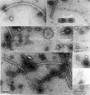
6. Chances are this is a new virus (or new subtype of virus) in the [family _Filoviridae_]. The only 2 known viruses in this group are Ebola and Marburg. It looks as though [the infection] may have entered Zambia from the Democratic Republic of the Congo (DRC) through a tick (carried on a horse), but again this cannot be confirmed.

This comment assumes that labs in South Africa have already tested all known VHFs. It is unlikely to be pneumonic plague, as this would have been discovered in South Africa; however, it is still a possibility that this [putative] viral disease has been in the Southern Province of Zambia (and that the 4 reported cases seen there were not diagnosed or wrongly called pneumonic plague).

7. The important steps in control are 1. effective quarantine of sick patients, and 2. monitoring of all “in-contact” cases, with quarantine as soon as any signs of flu or fever are noted. The government should also ideally make a statement to calm the panic and prevent people from fleeing the capital (potentially carrying the disease countrywide). This disease only spreads to people who are in very close contact with sick individuals. Those family members who are potentially incubating the disease should be encouraged to stay
around Lusaka, so that signs can be picked up quickly and treatment issued rapidly….[section on use of ribavirin – effective only against Lassa fever – edited out].

Communicated by:
Dr Amy Cantlay BVSc.MRCVS <inka@iwayafrica.com>
Veterinarian
Mkushi, Zambia

[ProMED-mail thanks Dr. Cantlay for her commentary, which contributes some interesting detail. At this point, it would not be useful to speculate further on the identity of the infectious agent responsible for the deaths of the 4 Zambian patients. No doubt a firm diagnosis will be available shortly from a South African reference laboratory. Several different viruses cause viral hemorrhagic fever. Of these, Ebola, Marburg, Lassa or Crimean-Congo hemorrhagic fever viruses have not been recorded in Zambia up to the present. A comprehensive account of these and other viruses responsible for hemorrhagic fevers can be found at the US CDC website: <http://www.cdc.gov/ncidod/diseases/virlfvr/virlfvr.htm>.

So: an unknown fever-causing agent, possibly associated with a tick bite in the index case, but which seems definitely to be transmitted quite efficiently via exposure to (presumably) body fluids, in a hospital setting…which does not appear to be known strains / types of Marburg or Ebola viruses, or Crimean-Congo haemorrhagic fever virus.

Loose in Johannesburg…part of a greater conurbation housing some 9 million people….

Should we be worried??

And the answer would be – NO.

If Ebola didn’t spread out of Kikwit in 1995 – a city of 500 000+ with no decent infrastructure to speak of – even to get as far as Kinshasa, then why should it spread in Johannesburg, or even Lusaka, where the infrastructure is MUCH more sophisticated?

I will leave this with a couple of quotes from posts I compiled on Ebola back in August 1995 from the fondly-remembered virology group at bio.net:

“To: virology@net.bio.net
From: ED@molbiol.uct.ac.za (“Ed Rybicki”)
Subject: Re: The Ebola virus – the end of the civilized world
Date: 18 Aug 1995 05:53:07 -0700

I would say you – and many others – are being unnecessarily
frightened by a concerted media campaign designed at selling lurid
books and films.  Listen – for a change – to what experts tell you,
and react accordingly.

That is, RELAX!!!!!”

And:

“To: virology@net.bio.net
From: york@mbcrr.harvard.edu (Ian A. York)
Subject: Re: Ebola: the greatest threat, continued

In article <99792FA2E69@ida.ruc.dk>,  wrote:
>
>Ebola is another ballgame. There is no way to protect effectively
>against this disease, and we have seen at mutation of this virus, Ebola
>Reston, that evidently was airborn (luckily it only affects monkeys).


Ebola has killed less than 400 people in the past decade.  By contrast, typhoid fever kills over 600,000 people per year; measles kills 1,000,000 (one million) people per year.  If you think Ebola has the potential tokill anywhere near that many, you don’t understand the virus.  The Ebola outbreak in Kikwit *was* the worst-case scenario; *everything* went wrong.  300 deaths.  Not trivial.  But a tiny fraction of the real killers. 

Lobby and try to get measles vaccine in Africa, if you want to do some good.  So don’t waste your time worrying about Ebola.”

Amen to that!  Pity we have to keep revisiting The Threat From Darkest Africa – maybe we can sell the rest of the world some vaccines against them sometime soon…B-)

HIV: roots run deeper than we knew

2 October, 2008

I have previously posted a number of articles on “molecular archaeology” of viruses, and how one can use extant sequences, archived tissue samples, or even blood of pandemic survivors to speculate on the origins of specific viruses, of viruses generally, or on the nature of old pandemic strains.Now HIV falls under the spotlight – again – as the 2nd October issue of Nature publishes three articles (one letter, a commentary on it and an independent commentary) on the origins of HIV-1 pandemic strains.I picked up on the first news – evidence for an older-than-previously-thought origin for HIV-1 – via our local paper this morning. Now this is VERY impressive; they usually keep science news for a slow day, and here they were telling us about a Nature paper on the day it was published! Accessing Nature brought up the Nature News commentary by Heidi Ledford, titled “Tissue sample suggests HIV has been infecting humans for a century”. Essentially, the commentary summarises the findings of Michael Worobey of the University of Arizona and his colleagues, who managed to amplify and sequence HIV-specific cDNA and DNA from a paraffin-embedded lymph node biopsy dating from 1960 from a woman in Léopoldville (now Kinshasa) in what is now the DR Congo. To quote Ledford:

“Their results showed that the most likely date for HIV’s emergence was about 1908, when Léopoldville was emerging as a centre for trade.”

Their findings added credibility to an earlier demonstration of HIV-1 in a 1959 sample, also from Kinshasa. What was interesting was that the sequences of the two viruses differed by 12%: this indicates that there was already significant divergence in the HIV-1 strains infecting people as early as 1960, pointing to a longer history of human infection than the previous estimate of the 1930s.Which led on to the Comments section, where one finds gems like this:

“This is one of the most stupid discovery I have ever heard. You will blame every single human plague on Africa, This is against all the Theories of evolutionary biology where The descents of the people that lived in the area might have developed a kind of resistance instead of being vulnerable to a new strain of the Virus.”

And:

“HIV is older than your great-grandparents, uh-huh! And I’ll bet that the US bio weapons effort is just ecstatic about this deflection. So now these members of science play to the bio-jackboot population controllers with this ‘revelation’ that those sex-crazed Africans of course just couldn’t stop themselves from pulling chimpanzees (I thought the original scientific theory was “green monkeys”) out of the trees for a quickie.”

I couldn’t take this, so I replied:

“It continues to amaze me, as a teacher of virology who tells big classes every year where HIV comes from, how every year some clique of students takes the African origin of HIV personally, as a direct affront. I echo the correspondent above: it is a virus, people. Viruses infect animals, they infect people, and sometimes spread from one to the other – and back, if you are a zoo animal and catch something from your handler. The AIDS pandemic is an accident of sociology, demography, access to high-speed, long-distance travel – and truck routes, and truck drivers. It happens that it originated in Africa. So did the human race – only a lot longer ago. Inevitably, as humans encroached on apes, things get passed across. And don’t spread, much, until…someone puts a road through the village.Why don’t people get more exercised about the origins of HTLV, another retrovirus that almost certainly jumped from monkeys to humans? Except that happened many thousands of years ago, and in south-east Asia, not Africa. And for the same reasons: people eat monkeys and great apes. For that matter, it is speculated that chimpanzees got SIV-CPZ from vervet (I HATE the term “green”) monkeys – and that it may have caused a population bottleneck, some 100 000 years ago. I note that chimpanzees are known to eat vervets, incidentally – so they caught the virus the same way we did.

Ah, well…. In any case, Paul Sharp of the University of Edinburgh – and phylogenetics guru – and the godmother of HIV/SIV diversity, Beatrice Hahn of the University of Alabama (from whom I got the chimp-vervet virus link), have an independent commentary in the same issue, wherein they speculate on “The prehistory of HIV-1”. They make this very interesting comment:

“If the epidemic grew roughly exponentially from only one or a few infected individuals around 1910 to the more than 55 million estimated to have been infected by 2007, there were probably only a few thousand HIV-infected individuals by 1960, all in central Africa. Given the diverse array of symptoms characteristic of AIDS, and the often-long asymptomatic period following infection, it is easy to imagine how the nascent epidemic went unrecognized.”

They also make the important point that the findings of the Worobey group were replicated – with similar but non-identical virus sequences being found – by another group working independently with the same tissue sample. This is important because it nails down the findings more firmly, as HIV sequences within an individual do differ, and:

“…the distance along the evolutionary tree from the group M ancestor to the ZR59 or DRC60 sequences is much shorter than those between the ancestor and modern strains, consistent with the earlier dates of isolation of ZR59 and DRC60, and confirming that these viruses are indeed old”.

a, The HIV-1 genome fragments that were successfully amplified from DRC60 (red) and are available for ZR59 (black). The numbering for the HIV-1 sequences corresponds to the HXB2 reference sequence (Supplementary Table 1). b, The A/A1 subtree from the unconstrained (in which a molecular clock is not enforced) BMCMC phylogenetic analysis. 1960.DRC60A is the University of Arizona consensus sequence, and 1960.DRC60N is the Northwestern University consensus sequence (that is, the sequences independently recovered in each of the two laboratories). The DRC60 sequences form a strongly supported clade with three modern sequences also sampled in the DRC.

Reproduced with permission from Nature Publishing Group (RightsLink License No 2041420001096) from:
Direct evidence of extensive diversity of HIV-1 in Kinshasa by 1960
Michael Worobey, Marlea Gemmel, Dirk E. Teuwen, Tamara Haselkorn, Kevin Kunstman, Michael Bunce, Jean-Jacques Muyembe, Jean-Marie M. Kabongo, Raphaël M. Kalengayi, Eric Van Marck, M. Thomas P. Gilbert & Steven M. Wolinsky
Nature 455, 661-664(2 October 2008) doi:10.1038/nature07390

So where did the virus infecting humans come from? The best guess, from the paper and the commentaries, is that it originated – as do the extant chimpanzee virus supposed to have descended from the common origin – in chimpanzees somewhere in southeast Cameroon.How did it get into people? Sharp and Hahn again:

“The simplest explanation for how SIV jumped to humans would be through exposure of humans to the blood of chimpanzees butchered locally for bushmeat.”

No sex, no weird practices…just eating our cousins.  And how and why did it get to Léopoldville? Trade…and in those days before widespread truck routes, that would have been via rivers – which, Sharp & Hahn point out, drain from southeast Cameroon into the Congo River, which flows past what is now Kinshasa. The Worobey paper has some interesting history in it, documenting times of founding and rates of growth of cities in equatorial west Africa: Léopoldville/Kinshasa was and probably still is by far the fastest-growing of these, and was the earliest founded (in 1885). All that was needed to seed a pandemic, then, was that people infected by a virus as a result of butchering chimpanzees, moved some 700 km down natural trade routes to an emergent trade centre – and settled, and passed it on.Then, of course, it is the same old story, told so well by Jared Diamond in “Guns, Germs and Steel“: increased human population density and breakdown in social structure leads to increase in rate of transmission and incidence / prevalence of a disease agent, until it reaches the threshold necessary to break out. It is interesting that it took so long to become noticed – but then, HIV is passed on considerably less efficiently than Hepatitis B virus, so the pace of the epidemic was necessarily slow.But very sure….

Big viruses have little viruses….

28 August, 2008

Just when you’d heard of mimiviruses, and thought it couldn’t get any stranger…the same team now bring you “mamavirus“, so named because it’s bigger!

But wait, that’s not all: apparently the new viruses have their very own “virophages” – smaller viruses which parasitise mamavirus-infected cells, and so called because they look like and have homology to bacteriophages.

And that’s still not all…Helen Pearson in the 6th August online Nature News then makes a case for viruses being considered as being alive, on the strength of this parasitism – and its detrimental effect on the larger virus, in terms of aberrant assembly, lower yield in infected cells, and so on.

http://www.nature.com/news/2008/080806/full/454677a.html

Well, now…some of us have never thought otherwise, have we?  And despite all of the hype about how huge these viruses are, and how they blur the boundary between alive and dead – they don’t do they?  For all their complexity, mimi- and presumbably mamaviruses do exactly what all other viruses do: they obligately parasitise cellular organisms, and use their machinery (and especially ribosomes) to make viral components which asemble into particles.

And the news piece goes on:

“The discovery of a giant virus that falls ill through infection by another virus is fuelling the debate about whether viruses are alive.

“There’s no doubt this is a living organism,” says Jean-Michel Claverie, a virologist at the the CNRS UPR laboratories in Marseilles, part of France’s basic-research agency. “The fact that it can get sick makes it more alive.””

Ye-e-e-ssss?  Really?  And calling what is obviously a satellite virus – for all that it is a big satellite virus – a “virophage” is simply creating new terms where none are necessary.  Actually, they go one worse than that: the original article refers to the satellite as “Sputnik” throughout, in a breathtaking display of artistic licence.

But putting the outraged taxonomist in me aside, this is a truly amazing discovery, worth all of the hype: it shows that we really don’t know a lot about what is sitting in plain sight – in cooling tower water, in this case – let alone what is is sitting in deep oceans, in terms of viral biodiversity.

While the mamavirus is interesting enough, what should be called Mamavirus associated satellite virus rather than Sputnik, is even more so: satellite viruses are generally small and have very few genes, whereas this has 21 genes in a ~18 kb circular dsDNA genome, makes isometric particles 50 nm in size (which can be found within mamavirus particles), and in the words of La Scola et al.:

“…contains genes that are linked to viruses infecting each of the three domains of life Eukarya, Archaea and Bacteria. Of the 21 predicted protein-coding genes, eight encode proteins with detectable homologues, including three proteins apparently derived from APMV [Acanthamoeba polyphaga mimivirus], a homologue of an archaeal virus integrase, a predicted primase–helicase, a packaging ATPase with homologues in bacteriophages and eukaryotic viruses, a distant homologue of bacterial insertion sequence transposase DNA-binding subunit, and a Zn-ribbon protein. The closest homologues of the last four of these proteins were detected in the Global Ocean Survey environmental data set, suggesting that Sputnik represents a currently unknown family of viruses.  Considering its functional analogy with bacteriophages, we classify this virus as a virophage. The virophage could be a vehicle mediating lateral gene transfer between giant viruses.”

Fascinating indeed: this parasite upon a parasite – it replicates only in the “giant virus factory found in amoebae co-infected with APMV” – is bigger than many autonomous viruses infecting mammals, looks like it is at least partly derived from a bacterial virus in that it may integrate into its host (the mimivirus?) within a eukaryote, and may shuffle DNA around between other viruses.

I’m definitely working in the wrong field.

There’s gold in them old veins….

26 August, 2008

I have often spoken of “molecular archeology” in my lectures, and of the possibility of identifying past epidemic / pandemic strains of human flu in particular, by looking at which viruses are recognised by antibodies from people who lived through the epidemics.

A new paper in Nature ups the stakes in this game considerably: a team led by one James E Crowe Jr describes how 32 survivors of the 1918 Spanish Flu pandemic – born in or before 1915 – were “mined” for antibodies, and seven donors additionally were shown to have circulating B cells which secreted antibodies which bound the 1918 H1N1 virus haemagglutinin (HA).  The team isolated 5 monoclonal antibodies from these subjects, and showed that these potently neutralised the infectivity of the virus and bound the HA of a 1930 swine virus, but did not cross-react with the HAs of more recent human  H1-containing viruses.

http://www.nature.com/nature/journal/vaop/ncurrent/abs/nature07231.html

Neutralizing antibodies derived from the B cells of 1918 influenza pandemic survivors : Abstract : Nature via kwout

 This achievement is undoubtedly a tour de force of modern molecular immunology – but is it useful?

Well, one very obvious fact is that people can obviously maintain significant levels of humoral immunity to viruses that infected them – in the words of the authors – “…well into the tenth decade of life.”  This is good news indeed for vaccinees who received vaccines for viruses which do not change much, like measles, mumps and poliomyelitis viruses.  However, given that influenza virus even of one H and N type can change so as to be unrecognisable in just a few years – the MAbs they generated did not react to any great extent with presumptively H1N1 human isolates from 1943, 1947, 1977 and 1999 – this is only of any use if the original virus were to be re-introduced somehow.

There was an intriguing statement in the paper which may shed some light on a long-running controversy as to the origin of the 1977 H1N1 pandemic, when the virus reappeared in humans for the first time since the early 1950s – allegedly as a result of an escape from a Soviet biowarfare lab.

The 1F1 antibody bound and neutralized the 1977 virus, albeit to a lesser degree than either the 1918 or the Sw/30 viruses … and to a minimal degree the 1943 virus”.

Ye-e-e-sssss…strange, that.  So the 1977 virus was antigenically more similar to 1930s era viruses than to one from 1943??

The proposed use of the findings also elicit biowar scenarios: for example, the fact that passive immunisation of people with antibodies to a particular virus can help them get over infection with it is purely academic for MAb to the 1918 virus – or is it?

I hope it is.

H5N1 flu: The End is still in sight

15 July, 2008

While the much-dreaded Big One – a major flu pandemic – still seems to be holding off, I am pleased to see that the latest edition of Nature has devoted an editorial and other commentary space to pandemic flu in general, and H5N1 flu in particular.

The Nature issue of 10th July 2008 – which has Ebola virus glycoprotein on the cover, about which, more later – has as its lead editorial title, “The long war against flu“.  The header goes on:

“That the H5N1 strain of bird flu has not yet caused a pandemic is no cause for complacency. Preparations for the inevitable must be redoubled to mitigate the potential devastation.”

My feelings entirely, and especially for the developing world – shared with you here and here, in MicrobiologyBytes, and here, in your own ViroBlogy.  It is all too easy to easy to fall into a state of complacency, or even H5N1 pandemic fatigue: however, this is really dangerous, especially for planners.  The editorial goes on:

“Five years after the deadly H5N1 avian influenza virus exploded into a global epidemic in birds, it has infected more than 300 people. Happily, it has not yet evolved into a strain that can transmit easily between humans — an event that would trigger a pandemic that could kill tens of millions. But as long as H5N1 continues to be present in animals, that risk persists. And with so many other flu strains out in the world, all constantly evolving, a flu pandemic is inevitable.”

 And:

“…improved control measures, especially for H5N1 itself, and public-health infrastructure are our frontline defences against a pandemic. Unfortunately, the overall control picture is bleak. Thailand, Vietnam and China have notched up successes in curbing outbreaks in birds, which is key to minimizing the chance that the virus can pass to humans. But South Korea had its worst outbreak ever in April, and the disease has become endemic in Indonesia, Bangladesh, Vietnam and Egypt. Eradication now seems impossible, and the task of containing the virus has become chronic and costly.”

Which sets the stage for Commentaries in the same issue: in “Ready for Avian Flu“, Tadataka Yamada et al. offer up “…a roadmap for heading off a global avian influenza catastrophe“.  They discuss how the WHO has made plans for stockpiling H5N1 vaccines, and that major vaccine manufacturers have offered to contribute – but also that the necessary allocation plan and ethics framework still need to be worked out.  They discuss how adjuvanting flu vaccines can reduce the dose by up to to four times, and how this together with dedicating the existing manufacturing framework – capable of some 500 million doses of trivalent vaccine a year  – to single-valency production, could allow some 6 billion doses a year to be produced.

Which, of course, would neatly cover the world’s population.  This sounds wonderful – but ignores the fact that H5N1 viruses are notoriously difficult to produce via conventional egg-based methods, which is what provides the bulk of the present manufacturing capacity….  Still, they also point out that adjuvanted, non-matched vaccines can still cross-protect against strains that have undergone seven or so years of genetic drift, meaning that stockpiled H5N1 vaccines could still be relevant in several years time.

They state the following:

“In the next 18 months we [Bill & Melinda Gates Foundation, the Pasteur Institute and the Wellcome Trust] will develop, maintain and disseminate a central inventory of funded research activities that are relevant to human influenza to ensure that stakeholders are well-informed. We will also coordinate roadmapping exercises to identify knowledge gaps. These will assist funders and researchers in establishing research-funding priorities, with specific focus on vaccines, drug therapies and epidemiology/population science (for example, diagnostics, surveillance, transmission and modelling). The Bill & Melinda Gates Foundation and the Wellcome Trust will collaborate to fund these activities.”

And how much developing country input will there be into this?  Distressingly little, probably, given the propensity of these funds to at best give money to developed country groups to work with developing country folk, but at least the roadmap addresses issues that are relevant to the whole world community.  Like intellectual property concerns, coordination of stockpiling and distribution, fair distribution, funding…and surveillance, the forgotten and possibly most important factor in determining if a flu outbreak is getting out of hand.  

Apropos of which, another commentary in the same issue – “The contents of the syringe“, by Steven Salzberg – notes that the influenza vaccine failed this northern hemisphere winter, and that future success relies on sharing data more widely and making the virus strain selection process more transparent.  Salzburg says:

 
“The WHO met on 11–13 February this year to decide on the strains to be included in the vaccine for the 2008–09 season. As usual, the meeting was closed to all but invited participants, who this year included members of the WHO influenza surveillance network, representatives of national drug regulatory agencies, and influenza vaccine manufacturers. The experts involved chose to replace the H3N2 strain with a more recent isolate, from 2007, which should be a better match to the circulating viruses next season. Neither the WHO nor the CDC publishes the evidence used to support their decision [my emphasis]. That evidence includes hemagglutinin inhibition tests of hundreds of isolates, genome sequences of some isolates and data on the ease with which the isolates can be grown in eggs.”

“The process of choosing flu-vaccine strains needs to be much more open. Other scientists, such as those in evolutionary biology with expertise in sequence analysis, could meaningfully contribute to the selection. At present, external scientists cannot review the data that went into the decision, nor can they suggest other types of data that might improve it.”

Whoops…so those who would safeguard us, feel no need to tell us what is going on??  Salzburg has some suggestions:

“The leaders of the influenza community, especially the WHO and the CDC, should create policies — for sharing data and isolates — that are more open, and should insist that their own scientists follow those policies. When these leading organizations set an example, the rest of the community will follow.”

“Of course, preparing vaccine in cell culture could reduce some of the pressures put on that dark room of vaccine predictors. The current system, in which most of the world’s vaccine supply is grown in chicken eggs, is an antiquated, inefficient method requiring six months or more to ramp up production, which in turn means that the vaccine strains must be chosen far in advance of each flu season. More crucially it sometimes prevents the use of the optimal strain, as it did in 2007. And, if the next pandemic is an avian-influenza strain such as H5N1, then it could easily sweep through the chicken farms that we rely on to produce eggs for vaccines. [me again]”

 He goes on to extol the benefits of sharing sequence information in particular, so as to enable rational, evidence-based choice of flu strains for vaccines – and the use of non-egg-based cell culture methods for vaccine production, and how these should allow far quicker development of flu vaccines.

All of this is very cogent and timeous.  However, it begs the question, previously raised in ViroBlogy, as to how production will be increased to take care of everyone who may be affected.

I still think plants are the answer…!  I note the plant-based flu vaccine group paper that I blogged on previously in this forum has a sequel: this is a better paper all around, and points up the need to explore this sort of production system for this kind of virus.

But I digress – so let me do so thoroughly.  The Nature issue also has a news item on the Eppendorf Song, a new piece of viral advertising with a boy band extolling the virtues of a multipettor.  Not a patch on the Biorad PCR Song, guys – but nice to see some popular culture making its way into science equipment advertising!

Viromics

25 June, 2008

Hot on the heels of the popular term genomics – defined by Wikipedia as “the study of an organism’s entire genome” – comes one for virologists: viromics.  There have been a number of articles in recent years on “viromes”, or the total viral genome content of the DNA found in certain biological sampling areas such as entire oceans or just in humans, so one might be forgiven for assuming that viromics was the study of entire viral genomes.

 But you might be wrong…for example, Applied Viromics of California have this to say:

“The term “viromics” was introduced on a biochemical pharmacology discussion conference in 2001 (Lotze MT and Kost TA. Cancer Gene Therapy. 2002 Aug; 9(8):692-9). It refers to “the use of viruses and viral gene transfer to explore the complexity arising from the vast array of new targets available from the human and murine genomes”.”

So: just about using viruses to study animals, then?  The Katze lab at the University of Washington – who I note have the word in their Web address – focus on:

…the use of genomic and proteomic technologies to study virus-host interactions and the varied strategies used by viruses to evade cellular defense mechanisms. We study a wide range of viral pathogens, including hepatitis C, influenza, Ebola, West Nile, SARS-associated coronavirus, herpes simplex virus, and human and simian immunodeficiency viruses.”

 So it’s studying human-virus interactions, then?  However, the Marine Microbiology Group at the Universrsity of South Florida has as its viromics interest

“…the interaction of viral genomes with their environments by sequencing several temperate/pseudotemperate marine phage genomes”.

 Much more like what I thought it was!  However, possibly the best definition I have seen is from a Nature Biotechnology Commentary on “Systems biology and the host response to viral infection“, which pops up the term “Systems virology” as a synonym for viromics.  the article states:

“There is increasing evidence from functional genomics experiments that the patterns of cellular response to a variety of viral infections may reflect the pathogenic properties of the viruses. We contend that dissection of the critical, and often subtly different, cellular pathways will eventually unveil opportunities for manipulating the host immune response to fight off viral infection, control pathogenesis or both.” 

And in a side box on viromics: 

“Innovations in sequencing technologies, particularly the rapid, high-throughput pyrosequencing platforms, continue to transform large-scale biology. … The fields of viral metagenomics and viral diagnostics are therefore poised for rapid expansion.”

Again – unsurprisingly, given that the Katze lab mentioned above is involved – a rather pathocentric approach, but all in all, a nice summary of current thinking.

So it is not the “what” so much as the “what it does” with viruses – at least, with the human virome.  Of course, for those of us with other interests, the new viromics can largely be concerned with discovery: there is a largely undescribed universe of viruses out there, in everything from seawater to duck ponds, biofilms to beehives, colons to parrots.  Cataloguing some of that diversity can only lead to new insights – which will lead to the kinds of questions that can be answered by “systems virology” approaches.

For instance, in my PhD thesis in 1984, I published an virus-like particles in plant extractselectron micrograph of a collection of unidentified particles found in preparations of known viruses, concentrated and partially purified from plant extracts.  there is a veritable zoo of things present, from a variety of fialments – some probably flagellae – to a motley collection of spheres and even some recognisable phages or parts thereof.  It has also been stuck on my office wall for the last ten years, since I found it kicking about in a drawer….

The point is, we generally find what we are looking for – and presume that what is present in the greatest abundance is just that, and then ignore everything else. 

Now we don’t have to do that any more: a metagenomic study of a particle-enriched preparation such as that seen on the right could yield fabulous riches, and unveil hitherto-unsuspected minor populations of plant viruses, unknown fungal and bacterial viruses, and even viruses infecting insects, which may be passively “vectored” by plants.

In a dramatic application of the worth of this sort of discovery, a major metagenomic project involving colony decline disorder of honeybees was described in MicrobiologyBytes recently: this involved sequencing all of the DNA and cDNA from many beehives, and doing a subtractive analysis to determine which possible pathogen was involved.

Other efforts under way in our laboratories in Cape Town include a study of the single-stranded circular DNA virome of grasses in Africa: this has resulted in six publications to date, and over 300 full mastrevirus genomic sequences in less than two years, and has radically altered our perception of both the diversity and the recombination potential of these commercially-important viruses.  This phenomenal progress has been enabled by recent technological breakthroughs – in this case, the use of bacteriophage phi29 DNA polymerase for isothermal “rolling circle” genome amplification, and cheap commercial sequencing.  This is rapidly diversifying into similar work on Beak and feather disease circoviruses in parrots, and even dsDNA viruses.

We are at the threshold of an era of significant discovery potential in virology, much as happened in the early 1990s with the advent of PCR – only more so.  The nice thing is that rapidly-developing sequencing techniques will allow ever-cheaper large-scale sequencing – so that finances will not be the limiting factor they may have been up to now, and developing countries can share in the viromic bounty.

And every now and then, the young people around will tell me what is going on – for which I am grateful.

West Nile virus vaccine: almost a replicant

2 June, 2008

West Nile virus – a member of the family Flaviviridae – has insidiously spread halfway around the world from its origins in Africa, in just a few years.  It invaded the east coast of the USA, probably from the Middle East,  via either infected birds, mosquitoes, humans, or another vertebrate host in around 1999; since then it has spread all the way across the continent to the west coast, and has become truly endemic. 

Virions have a regular icosahedral-type structure, despite being enveloped, as a result of a structured nucleocapsid and a highly-structured array of envelope glycoprotein.  They contain a positive strand RNA genome of ~11 kb with a single long open reading frame that is translated as a polyprotein of about 3400 amino acids, which is then processed into individual regulatory and structural proteins.

The virus subtype spreading in North America – lineage 1 – causes encephalitis in humans, unlike the enzootic variant circulating in birds and animals in Africa.  It also cause severe mortality – near 100% in experimentally infected animals – among American Crows and other corvids: a feature of the spread of the disease has been dead crows found in and around towns in the USA.  A feature of lineage 1 viruses is their infection of horses and other equines as well – with up to one in three clinically-infected horses dying.  The human impact, however, is seen as a major problem: systemic febrile illness develops in ~20% of those infected with WNV, while severe neurologic illness developes in <1% of persons infected – with mortality rates of 5 -14% among persons with neurologic symptoms in recent US, Romanian, Russian, and Israeli outbreaks.

There has been a concentrated effort to develop a human vaccine or vaccines since the onset of the US epidemic – horse vaccines are already commercially available – and our knowledge of the virus has benefitted greatly as a result.  This includes a detailed structure for the virus, obtained by cryoelectron microscopy image reconstruction.

http://www.purdue.edu/uns/html4ever/031009.Kuhn.westnile.html

Purdue team solves structure of West Nile virus via kwout

 Now a team led by Alexander Khromykh from Brisbane in Queensland, Australia, writing in the May issue of Nature Biotechnology, have described a novel “single-round infectious particle” DNA vaccine against WNV which significantly increases protection in mice to lethal challenge with the live virus.   In the words of the authors:

“We augment the protective capacity of a capsid-deleted flavivirus DNA vaccine by co-expressing the capsid protein from a separate promoter. In transfected cells, the capsid-deleted RNA transcript is replicated and translated to produce secreted virus-like particles lacking the nucleocapsid. This RNA is also packaged with the help of co-expressed capsid protein to form secreted single-round infectious particles (SRIPs) that deliver the RNA into neighboring cells. In SRIP-infected cells, the RNA is replicated again and produces additional virus-like particles, but in the absence of capsid RNA no SRIPs are formed and no further spread occurs. Compared with an otherwise identical construct that does not encode capsid, our vaccine offers better protection to mice after lethal West Nile virus infection. It also elicits virus-neutralizing antibodies in horses. This approach may enable vaccination against pathogenic flaviviruses other than West Nile virus.”

Adapted by permission from Macmillan Publishers Ltd: Nature Biotechnology 26, 571 – 577, 20 April 2008 doi:10.1038/nbt1400 Single-round infectious particles enhance immunogenicity of a DNA vaccine against West Nile virus, David C Chang et al., copyright 2008

This is a very clever use of fundamental knowledge of virus structure and assembly: the virus envelope proteins – E and prM – can form budded particles if expressed in isolation; if expressed with the capsid protein, the particles encapsidate RNA with the appropriate encapsidation signal to form virions.  The DNA vaccine encodes a transcriptional unit corresponding to a viral genome which lacks only the capsid protein gene, as well as a separate capsid gene under back-to-back cytomegalovirus (CMV) promoters.  Thus, cells transfected with the DNA vaccine can produce both virus-like prM and E protein and membrane particles (VLPs), or pseudovirions which in addition contain a capsid and the engineered (=lacking capsid protein gene) genome.  While both are highly immunogenic, the pseudovirions can additionally infect other cells to release replicative genomic RNA, which can produce VLPs but not pseudovirions, as the capsid protein-encoding RNA is not encapsidated.  Thus, initial transfection leads to release of particles which allow a single subsequent round of VLP production, but no further spread of the replicative RNA.

A very clever trick – and worthy of being repeated for a number of related pathogenic flaviruses, including dengue and yellow fever viruses.

Even if the particles can’t pass the Voight-Kampff test…B-)