Archive for the ‘Viruses’ Category

Is This The Big One?

28 April, 2009

28th April 2009:

It just HAD to happen.

There was the world’s attention, focussed on H5N1 bird flu from Asia as The Next Big One – including doom and gloom pronouncements from right here (and here) – and of course, another flu comes from another source, in another location entirely.  You can, however, as previously highlighted here in ViroBlogy, use Google “Flu Trends” to track it – and now Google Maps too (thanks, Vernon!).

Flu life cycle

Flu life cycle

So what do we know?  On the 27th of  April, the Mexican government admitted to some 150 deaths, and over 1600 people apparently infected, in an epidemic caused by an Influenza A H1N1 virus that appeared to be a reassortant of viruses from pigs, birds and humans.  The virus has been dubbed “swine flu”; however, there is doubt as to whether it has been shown to even infect pigs, let alone been found in them, and it probably ought to be known as “Mexico Flu”.  There is the problem, of course, that apparently parts of the virus – and the N1 gene in particular – are of Eurasian swine flu origin, so exactly where it comes from may be forever obscure.

 As for current expert knowledge, the Centers for Disease Control and Prevention (CDC) and the World Health Organisation (WHO) have set up dedicated pages to track the potential pandemic – because that is what they are calling it.

The WHO has, as of the 27th April,

“…raised the level of influenza pandemic alert from the current phase 3 to phase 4.

http://blogs.wsj.com/health/2009/04/27/understanding-the-whos-global-pandemic-alert-levels/

Swine Flu: Understanding the WHO’s Global Pandemic-Alert Levels – Health Blog – WSJ via kwout

The change to a higher phase of pandemic alert indicates that the likelihood of a pandemic has increased, but not that a pandemic is inevitable.

As further information becomes available, WHO may decide to either revert to phase 3 or raise the level of alert to another phase.

This decision was based primarily on epidemiological data demonstrating human-to-human transmission and the ability of the virus to cause community-level outbreaks.

Given the widespread presence of the virus, the Director-General considered that containment of the outbreak is not feasible. The current focus should be on mitigation measures.”

All of which begs the questions: what IS it, and how BAD is it??  We know that by the 28th April, the virus had been confirmed in the USA (>40 cases), Spain, Canada, and according the the BBC, the UK, Brazil and New Zealand as well.

While financial markets are panicking , airlines are cancelling flights, and people in Mexico appear to be dying, people infected in the USA seem only to be getting ill, and then recovering.

The bad news is that the virus haemagglutinin – the H1 – is probably only distantly related to that of the currently circulating human variant, so the flu vaccines on release right now will be of only limited efficacy.

The good news – especially for Roche and GlaxoSmithKline – is that the antivirals Tamiflu and Relenza seem to work against the virus.

29th April 2009

The virus continues to spread: according to the WHO site,

“As of 19:15 GMT, 28 April 2009, seven countries have officially reported cases of swine influenza A/H1N1 infection. The United States Government has reported 64 laboratory confirmed human cases, with no deaths. Mexico has reported 26 confirmed human cases of infection including seven deaths. The following countries have reported laboratory confirmed cases with no deaths – Canada (6), New Zealand (3), the United Kingdom (2), Israel (2) and Spain (2).
….
WHO advises no restriction of regular travel or closure of borders. It is considered prudent for people who are ill to delay international travel and for people developing symptoms following international travel to seek medical attention, in line with guidance from national authorities.

There is also no risk of infection from this virus from consumption of well-cooked pork and pork products. Individuals are advised to wash hands thoroughly with soap and water on a regular basis and should seek medical attention if they develop any symptoms of influenza-like illness.

Of course, there is also the inevitable hype – and some humour (thanks, Suhail!):

With a byline reminiscent of the “Ebola Preston” which was coined to satirise the hype generated around the 1995 Ebola hype, we have

 30th April 2009:

…so of course, I talk to a journalist; and of course, I shouldn’t have…!  For an otherwise good article about pandemic preparedness in Africa [ignore the bit about no drug stockpile in South Africa, because apparently we have some], see here.  South Africans: look at info on influenza at the National Institute for Communicable Diseases (NICD) in Johannesburg.

The WHO yesterday raised the level of influenza pandemic alert from the current phase 4 to phase 5.  We owe the WHO Director-General, Dr Margaret Chan, for these comments:

On the positive side, the world is better prepared for an influenza pandemic than at any time in history.

Preparedness measures undertaken because of the threat from H5N1 avian influenza were an investment, and we are now benefitting from this investment.

For the first time in history, we can track the evolution of a pandemic in real-time.”

From the BBC today:

In Mexico, the epicentre of the outbreak, the number of confirmed cases rose to 97 – up from 26 on Wednesday….

  • The Netherlands confirms its first case of swine flu, in a three-year-old boy recently returned from Mexico. Cases have also been confirmed in Switzerland, Costa Rica and Peru
  • The number of confirmed cases in the US rose to 109 in 11 states
  • Japan reported its first suspected case of swine flu
  • China’s health minister says that the country’s scientists have developed a “sensitive and fast” test for spotting swine flu in conjunction with US scientists and the WHO. The country has recorded no incidence of the flu yet.
  • The WHO says it will now call the virus influenza A (H1N1).

And first prize for over-reaction of the year:

On Wednesday, Egypt began a mass slaughter of its pigs – even though the WHO says the virus was now being transmitted from human to human [and there is no evidence it was ever transmitted between pigs].


Index: ViroBlogy / MicrobiologyBytes flu-related posts

Umm…no, they don’t

28 November, 2008

For shame, New Scientist!!  There I go recommending you to all and sundry – and especially my students – as a fount of general scientific knowledge, and you do this!!  In the 22nd November issue – freshly on my desk, down here in the Deepest South – there is, under the column heading “60 Seconds”, the following snippet:

Shot thwarts warts

A human papilloma virus vaccine already approved in women to prevent cervical cancer has proved equally effective in men against genital wartswhich can lead to cervical cancer in women. In a trial of the vaccine in 4000 men only three recipients developed HPV-related lesions compared with 31 who received a placebo.

Note the bolded section: BECAUSE IT IS VERY WRONG. 

No, New Scientist, genital warts in men do NOT lead to cervical cancer in women: warts in men and women are caused by a number of what are termed “low risk” viruses (for low cancer risk), and especially by HPV types 6 and 11 – which are two of the types included in Merck’s Gardasil.  The other two types in this vaccine – which are the same ones as in GlaxoSmithKline’s Cervarix – are the high cancer risk HPV types 16 and 18.  Which do NOT cause genital warts, in men or women: rather, they cause inapparent infections of the epithelial tissue of the penis in men, and of the vaginal and cervical mucosa in women. 

The lesions can most often only be seen in both men and women after they have been painted with an acetic acid solution – hence the name “aceto-white lesion”.

The fact that warts are apparently prevented in men is a very welcome development: this means that in all likelihood the other infections will be prevented too, and men will not be able to transmit HPV 16 and 18 to women.

Which is presumably what you meant.

Despair

25 November, 2008

I have had much occasion to frequent the archives of Despair.com in search of demotivational posters with which to inspire my friends and colleagues…and now Alan Cann has given me the opening to display one of my better efforts.  He posted on a new Ebola virus found recently in western Uganda.

I will give you this….

ebola-poster1

Zambia fever virus: latest

8 October, 2008

Latest links:

120 people under observation for killer virus” SABC, October 8th

“Killer-fever link found by luck ” Independent On-Line 08-10-08

And just as a reminder that life goes on: a Congo fever patient from here in South Africa.

Deadly Zambian fever: deja vu all over again

7 October, 2008

The news media are presently fascinated by the appearance of what looks like a new and nasty virus from my old home country, Zambia: see a link to The Times article of 7th October for the official word.

Which is “Don’t Panic”, written in large, friendly letters across the face of the newspaper….

From The Times article by Sashni Pather:

Disease transmitted via bodily fluids

THE deputy director of the National Institute for Communicable Diseases has assured the public that there is no need to panic, despite the fact that four people have been killed by an unknown, highly contagious virus.

Doctor Lucille Blumberg, who also heads up the NICD’s epidemiology unit and consults to its special pathogens unit, referred to the death of Cecilia van Deventer, 36, as an “isolated case” and said test results were not yet available.

…A paramedic, Hannes Els, 33, who treated the critically ill Van Deventer in Zambia, and brought her to the Morningside Medi-Clinic in Sandton on September 12, died last Thursday after being infected with the highly contagious disease.

On Sunday a nurse and a cleaner, who had possibly been exposed to the disease, died.

Blumberg said: “The cause of death of the cleaner is still being investigated. We are busy conducting tests on all four deceased. The cleaner might not have been killed as a result of the virus.

“This is an isolated case. There have been no other reported cases in Lusaka, Zambia. “”

OK, so no obvious panic there, then – but disturbing echoes of another incident from 1996, when a Gabonese doctor was medevacced in to South Africa, and passed on an Ebola virus infection to a theatre nurse, Marilyn Lahana.  He recovered, and she died – and there was close to panic in the land, as chronicled here.  And here we are again….  Incidentally, anyone who wants to see how the first Ebola outbreaks of the electronic age unfolded can see a day-by-day history here, on my original Ebola pages.  Still accessible, to my surprise!

That wonderful institution that is ProMED – who were the first people to break hard news of the Kikwit Ebola outbreak back in 1995, came to the attention of the serious medical reporting world as a result – has a slightly different view of the whole thing – and some interesting details not in the general news story.  In this morning’s digest:

UNDIAGNOSED FATALITIES – SOUTH AFRICA ex ZAMBIA (02)
***********************************************
A ProMED-mail post
<http://www.promedmail.org>
ProMED-mail is a program of the International Society for Infectious Diseases
<http://www.isid.org>

[1]
Date: Mon 6 Oct 2008
Source: South African Broadcasting Corporation News online [edited]
<http://www.sabcnews.com/south_africa/health/0,2172,177844,00.htm>

A 4th person with viral haemorragic fever (VHF) symptoms has died. The virus has already claimed the lives of a Zambian national and 2 other people at the Morningside Clinic in Johannesburg. The woman was a cleaner at the clinic.

The National Health Department has issued an alert in Gauteng following these deaths. Unconfirmed tests indicate they may have died of [a] fatal viral haemorragic fever. An Outbreak Response and Tracking Team has been set up to contain any further spread. The department’s Zanele Mngadi says investigations are still underway into the cause of the deaths.

Mngadi confirmed the death of the 4th person, who was admitted at the Leratong hospital last night [5 Oct 2008]. The patient, who showed symptoms of VHF, was transferred to the Charlotte Maxeke Johannesburg
Academic Hospital, where she died. The health department says there is no need for South Africans to panic. The department’s Frew Denson says the fever is highly contagious but is only transmitted through body fluids.

It is reported that the virus can kill a person within 72 hours. VHF is an extremely infectious and life-threatening disease caused by [several different] viruses, including Ebola virus. The death rate [in the case of Ebola virus] can be as high as 90 percent. Symptoms vary but include fever, vomiting, diarrhea and bleeding.

Communicated by:
Rabelani Daswa <rabedaswa@gmail.com>

******
[2]
Date: Mon 6 Oct 2008
From: Amy Cantlay <inka@iwayafrica.com>

I have just read the posting (Undiagnosed fatalities – South Africa ex Zambia: RFI 20081005.3139) on your site, and it appears to be rather misleading. The chronological order of events (as I can gather) is as follows (None of this information has yet been confirmed.):

4 Sep 2008 – Index Case – female South African, (living in Zambia for many years) begins to suffer from flu-like symptoms.

9 Sep 2008 – She is slowly deteriorating. She sees multiple doctors in Lusaka.

11 Sep 2008 – She is admitted to hospital and deteriorates over night.

12 Sep 2008 – Paramedic is called in to evacuate her to South Africa.  He does the transfer, along with another Dr assisting.

13 Sep 2008 – Index Case dies.

14 Sep 2008 Paramedic starts to develop flu-like symptoms.

14-27 Sep 2008 – Paramedic slowly deteriorates.

27 Sep 2008 – Paramedic is diagnosed as very sick and medivaced [sic] to South Africa. Nurse who treated Index case begins to get flu-like symptoms.

30 Sep 2008 – Paramedic dies.

1 Oct 2008 – Nurse who treated Index Case is admitted to hospital.

5 Oct 2008 – Nurse who treated Index Case dies.

The information that I can gather is the following:

1. Incubation period is as little as 2 days (paramedic), but as long as 14 days (nurse).

2. Disease course is generally 4-7 days of flu-like illness with patient only becoming critically ill in 2nd week of disease.

3. Further information is that Index Case reportedly had an eschar on one of her feet, thought to be from a tick-bite. She had also been in contact with horses from Congo in the weeks preceding her illness. Transmission is hypothesized to be by 2 means: tick-borne 1st (which may have brought the disease into the human population from the animal population) followed by direct contact with bodily fluids (resulting in human to human transmission).

4. It appears further hospital staff are now critically ill in Zambia, though this has not been confirmed.

5. If the incubation period is as long as 2 weeks, then we should still be closely watching all “contact-cases” for any signs of the disease. Those in contact with the Index case should be in the clear by now, while those in contact with the paramedic and the nurse (as well as any hospital staff who are currently sick) are still at high risk. One should probably work on a 21-day incubation period/quarantine period to be safe.

6. Chances are this is a new virus (or new subtype of virus) in the [family _Filoviridae_]. The only 2 known viruses in this group are Ebola and Marburg. It looks as though [the infection] may have entered Zambia from the Democratic Republic of the Congo (DRC) through a tick (carried on a horse), but again this cannot be confirmed.

This comment assumes that labs in South Africa have already tested all known VHFs. It is unlikely to be pneumonic plague, as this would have been discovered in South Africa; however, it is still a possibility that this [putative] viral disease has been in the Southern Province of Zambia (and that the 4 reported cases seen there were not diagnosed or wrongly called pneumonic plague).

7. The important steps in control are 1. effective quarantine of sick patients, and 2. monitoring of all “in-contact” cases, with quarantine as soon as any signs of flu or fever are noted. The government should also ideally make a statement to calm the panic and prevent people from fleeing the capital (potentially carrying the disease countrywide). This disease only spreads to people who are in very close contact with sick individuals. Those family members who are potentially incubating the disease should be encouraged to stay
around Lusaka, so that signs can be picked up quickly and treatment issued rapidly….[section on use of ribavirin – effective only against Lassa fever – edited out].

Communicated by:
Dr Amy Cantlay BVSc.MRCVS <inka@iwayafrica.com>
Veterinarian
Mkushi, Zambia

[ProMED-mail thanks Dr. Cantlay for her commentary, which contributes some interesting detail. At this point, it would not be useful to speculate further on the identity of the infectious agent responsible for the deaths of the 4 Zambian patients. No doubt a firm diagnosis will be available shortly from a South African reference laboratory. Several different viruses cause viral hemorrhagic fever. Of these, Ebola, Marburg, Lassa or Crimean-Congo hemorrhagic fever viruses have not been recorded in Zambia up to the present. A comprehensive account of these and other viruses responsible for hemorrhagic fevers can be found at the US CDC website: <http://www.cdc.gov/ncidod/diseases/virlfvr/virlfvr.htm>.

So: an unknown fever-causing agent, possibly associated with a tick bite in the index case, but which seems definitely to be transmitted quite efficiently via exposure to (presumably) body fluids, in a hospital setting…which does not appear to be known strains / types of Marburg or Ebola viruses, or Crimean-Congo haemorrhagic fever virus.

Loose in Johannesburg…part of a greater conurbation housing some 9 million people….

Should we be worried??

And the answer would be – NO.

If Ebola didn’t spread out of Kikwit in 1995 – a city of 500 000+ with no decent infrastructure to speak of – even to get as far as Kinshasa, then why should it spread in Johannesburg, or even Lusaka, where the infrastructure is MUCH more sophisticated?

I will leave this with a couple of quotes from posts I compiled on Ebola back in August 1995 from the fondly-remembered virology group at bio.net:

“To: virology@net.bio.net
From: ED@molbiol.uct.ac.za (“Ed Rybicki”)
Subject: Re: The Ebola virus – the end of the civilized world
Date: 18 Aug 1995 05:53:07 -0700

I would say you – and many others – are being unnecessarily
frightened by a concerted media campaign designed at selling lurid
books and films.  Listen – for a change – to what experts tell you,
and react accordingly.

That is, RELAX!!!!!”

And:

“To: virology@net.bio.net
From: york@mbcrr.harvard.edu (Ian A. York)
Subject: Re: Ebola: the greatest threat, continued

In article <99792FA2E69@ida.ruc.dk>,  wrote:
>
>Ebola is another ballgame. There is no way to protect effectively
>against this disease, and we have seen at mutation of this virus, Ebola
>Reston, that evidently was airborn (luckily it only affects monkeys).


Ebola has killed less than 400 people in the past decade.  By contrast, typhoid fever kills over 600,000 people per year; measles kills 1,000,000 (one million) people per year.  If you think Ebola has the potential tokill anywhere near that many, you don’t understand the virus.  The Ebola outbreak in Kikwit *was* the worst-case scenario; *everything* went wrong.  300 deaths.  Not trivial.  But a tiny fraction of the real killers. 

Lobby and try to get measles vaccine in Africa, if you want to do some good.  So don’t waste your time worrying about Ebola.”

Amen to that!  Pity we have to keep revisiting The Threat From Darkest Africa – maybe we can sell the rest of the world some vaccines against them sometime soon…B-)

There’s gold in them old veins….

26 August, 2008

I have often spoken of “molecular archeology” in my lectures, and of the possibility of identifying past epidemic / pandemic strains of human flu in particular, by looking at which viruses are recognised by antibodies from people who lived through the epidemics.

A new paper in Nature ups the stakes in this game considerably: a team led by one James E Crowe Jr describes how 32 survivors of the 1918 Spanish Flu pandemic – born in or before 1915 – were “mined” for antibodies, and seven donors additionally were shown to have circulating B cells which secreted antibodies which bound the 1918 H1N1 virus haemagglutinin (HA).  The team isolated 5 monoclonal antibodies from these subjects, and showed that these potently neutralised the infectivity of the virus and bound the HA of a 1930 swine virus, but did not cross-react with the HAs of more recent human  H1-containing viruses.

http://www.nature.com/nature/journal/vaop/ncurrent/abs/nature07231.html

Neutralizing antibodies derived from the B cells of 1918 influenza pandemic survivors : Abstract : Nature via kwout

 This achievement is undoubtedly a tour de force of modern molecular immunology – but is it useful?

Well, one very obvious fact is that people can obviously maintain significant levels of humoral immunity to viruses that infected them – in the words of the authors – “…well into the tenth decade of life.”  This is good news indeed for vaccinees who received vaccines for viruses which do not change much, like measles, mumps and poliomyelitis viruses.  However, given that influenza virus even of one H and N type can change so as to be unrecognisable in just a few years – the MAbs they generated did not react to any great extent with presumptively H1N1 human isolates from 1943, 1947, 1977 and 1999 – this is only of any use if the original virus were to be re-introduced somehow.

There was an intriguing statement in the paper which may shed some light on a long-running controversy as to the origin of the 1977 H1N1 pandemic, when the virus reappeared in humans for the first time since the early 1950s – allegedly as a result of an escape from a Soviet biowarfare lab.

The 1F1 antibody bound and neutralized the 1977 virus, albeit to a lesser degree than either the 1918 or the Sw/30 viruses … and to a minimal degree the 1943 virus”.

Ye-e-e-sssss…strange, that.  So the 1977 virus was antigenically more similar to 1930s era viruses than to one from 1943??

The proposed use of the findings also elicit biowar scenarios: for example, the fact that passive immunisation of people with antibodies to a particular virus can help them get over infection with it is purely academic for MAb to the 1918 virus – or is it?

I hope it is.

PIPO, I see you…

30 May, 2008

Now here’s an interesting thing: a completely unsuspected gene – as in, on open reading frame (ORF) that actually DOES something – in one of the best-studied familes of plant viruses.  From the International Service for the Acquisition of Agribiotech Applications (ISAAA)’s CropBiotech Update 30 May 2008:

Scientists Discover Hidden Gene in Major Plant Virus Family

The virus family Potyviridae includes more than 30 percent of known plant virus species, most of which are of great agricultural significance such as the potato virus Y, turnip mosaic virus and wheat streak mosaic virus. Scientists from the Iowa State University, working with colleagues from the University College Cork in Ireland, have discovered a tiny gene present in all members of this virus family. Without this gene, the viruses are harmless.

Using a gene-finding software, the team identified a stretch of nucleotide bases that overlaps with a much larger and well characterized gene in potyviruses. They called the new gene pipo (short for pretty interesting potyvirus ORF). Alterations in the sequence of the pipo gene, while leaving the polyprotein amino acid sequence unaltered, were found to be lethal for the viruses.

The team led by Allen Miller and John Atkins are now working to determine the function of gene during infection as well as how the pipo protein is expressed from the viral genome. For this, the U.S. Department of Agriculture National Research Initiative (USDA-NRI) has awarded them with a $400,000 competitive grant.

For more information, visit \http://www.public.iastate.edu/~nscentral/ Read the paper published by PNAS at http://www.pnas.org/cgi/reprint/105/15/5897

Nice one, guys…$400 000 should buy a few more ORFs…B-)  Seriously, though, the dogma has been for years that potyviruses, like picornaviruses, have a single long (~10kb) ORF, which expresses a polypeptide from the genomic RNA which is cotranslationally processed into a number of different proteins – and that was all there was.  This discovery is like finding a new and secret drawer in an old and familiar chest of drawers, or an extra pocket in your trousers.  Or, as I did recently, that there wer two interior lights in my car which I had not known of for six years…but I digress.

In the words of the authors:

“We report the discovery of a short ORF embedded within the P3 cistron of the polyprotein but translated in the +2 reading-frame. The ORF, termed pipo, is conserved and has a strong bioinformatic coding signature throughout the large and diverse Potyviridae family. Mutations that knock out expression of the PIPO protein in Turnip mosaic potyvirus but leave the polyprotein amino acid sequence unaltered are lethal to the virus. Immunoblotting with antisera raised against two nonoverlapping 14-aa antigens, derived from the PIPO amino acid sequence, reveals the expression of an ~25-kDa PIPO fusion product in planta. This is consistent with expression of PIPO as a P3-PIPO fusion product via ribosomal frameshifting or transcriptional slippage at a highly conserved G1-2A6-7 motif at the 5′ end of pipo. This discovery suggests that other short overlapping genes may remain hidden even in well studied virus genomes (as well as cellular organisms)…”

They go on to tout the virtues of the “software package MLOGD”, which it turns out is from here (Firth AE, Brown CM (2006) Detecting overlapping coding sequences in virus genomes. BMC Bioinformatics 7:75), and is the Maximum Likelihood Overlapping Gene Detector.   They say:

“Tests show that, from an alignment with just 20 mutations, MLOGD can discriminate non-overlapping CDSs from non-coding ORFs with a typical accuracy of up to 98%, and can detect CDSs overlapping known CDSs with a typical accuracy of 90%. In addition, the software produces a variety of statistics and graphics, useful for analysing an input multiple sequence alignment.”

And yes, it does make nice pictures: see this and this for examples.

All of which simply goes to reinforce my conviction that virus genomes may be generally quite small, but small does not necessarily mean simple.  Small means having to compress information, reuse sequences – and overlap ORFs in unsuspected ways.

Oxygen from viruses??

7 April, 2008

I thank my colleague Suhail Rafudeen for alerting me to this:

 “Some Of Our Oxygen Is Produced By Viruses Infecting Micro-organisms In The Oceans

ScienceDaily (Apr. 6, 2008) – Some of the oxygen we breathe today is being produced because of viruses infecting micro-organisms in the world’s oceans, scientists heard April 2, 2008 at the Society for General Microbiology’s 162nd meeting.

About half the world’s oxygen is being produced by tiny photosynthesising creatures called phytoplankton in the major oceans. These organisms are also responsible for removing carbon dioxide from our atmosphere and locking it away in their bodies, which sink to the bottom of the ocean when they die, removing it forever and limiting global warming.

“In major parts of the oceans, the micro-organisms responsible for providing oxygen and locking away carbon dioxide are actually single celled bacteria called cyanobacteria,” says Professor Nicholas Mann of the University of Warwick. “These organisms, which are so important for making our planet inhabitable, are attacked and infected by a range of different types of viruses.”

The researchers have identified the genetic codes of these viruses using molecular techniques and discovered that some of them are responsible for providing the genetic material that codes for key components of photosynthesis machinery.

“It is beginning to become to clear to us that at least a proportion of the oxygen we breathe is a by-product of the bacteria suffering from a virus infection,” says Professor Mann. “Instead of being viewed solely as evolutionary bad guys, causing diseases, viruses appear to be of central importance in the planetary process. In fact they may be essential to our survival.”

Viruses may also help to spread useful genes for photosynthesis from one strain of bacteria to another.

Adapted from materials provided by Society for General Microbiology, via EurekAlert!, a service of AAAS”

Fascinating concept: viruses as an essential link in the circle of life?!  Not so far-fetched, though: just because we know them largely because of their propensity to cause, and our fascination with, diseases that affect us and our livestock and crops…doesn’t mean that is all there is.

Viruses have been around as long as any other form of life, and it would be strange indeed if some form(s) of commensalism and/or symbiosis had not evolved.

…and see here for some fascinating speculations on the possible involvement of viruses with the origin of eukaryotes.

Virus origins: from what did viruses evolve or how did they initially arise?

19 March, 2008

This was originally written as an Answer to a Question posted to Scientific American Online; however, as what they published was considerably shorter and simpler than what I wrote, I shall post the [now updated] original here.

The answer to this question is not simple, because, while viruses all share the characteristics of being obligate intracellular parasites which use host cell machinery to make their components which then self-assemble to make particles which contain their genomes, they most definitely do not have a single origin, and indeed their origins may be spread out over a considerable period of geological and evolutionary time.

Viruses infect all types of cellular organisms, from Bacteria through Archaea to Eukarya; from E. coli to mushrooms; from amoebae to human beings – and virus particles may even be the single most abundant and varied organisms on the planet, given their abundance in all the waters of all the seas of planet Earth.  Given this diversity and abundance, and the propensity of viruses to swap and share successful modules between very different lineages and to pick up bits of genome from their hosts, it is very difficult to speculate sensibly on their deep origins – but I shall outline some of the probable evolutionary scenarios.

The graphic depicts a possible scenario for the evolution of viruses: “wild” genetic elements could have escaped, or even been the agents for transfer of genetic information between, both RNA-containing and DNA-containing “protocells”, to provide the precursors of retroelements and of RNA and DNA viruses.  Later escapes from Bacteria, Archaea and their progeny Eukarya would complete the virus zoo.

virus descent

It is generally accepted that many viruses have their origins as “escapees” from cells; rogue bits of nucleic acid that have taken the autonomy already characteristic of certain cellular genome components to a new level.  Simple RNA viruses are a good example of these: their genetic structure is far too simple for them to be degenerate cells; indeed, many resemble renegade messenger RNAs in their simplicity.

RdRp cassettes and virus evolution

RNA virus supergroups and RdRp and CP cassettes

What they have in common is a strategy which involves use of a virus-encoded RNA-dependent RNA polymerase (RdRp) or replicase to replicate RNA genomes – a process which does not occur in cells, although most eukaryotes so far investigated do have RdRp-like enzymes involved in regulation of gene expression and resistance to viruses.  The surmise is that in some instances, an RdRp-encoding element could have became autonomous – or independent of DNA – by encoding its own replicase, and then acquired structural protein-encoding sequences by recombination, to become wholly autonomous and potentially infectious.

A useful example is the viruses sometimes referred to as the “Picornavirus-like” and “Sindbis virus-like” supergroups of ssRNA+ viruses, respectively.  These two sets of viruses can be neatly divided into two groups according to their RdRp affinities, which determine how they replicate.  However, they can also be divided according to their capsid protein affinities, which is where it is obvious that the phenomenon the late Rob Goldbach termed “cassette evolution” has occurred: some viruses that are relatively closely related in terms of RdRp and other non-structural protein sequences have completely different capsid proteins and particle morphologies, due to acquisition by the same RdRp module of different structural protein modules.

Given the very significant diversity in these sorts of viruses, it is quite possible that this has happened a number of times in the evolution of cellular organisms on this planet – and that some single-stranded RNA viruses like bacterial RNA viruses or bacteriophages and some plant viruses (like Tobacco mosaic virus, TMV) may be very ancient indeed.

However, other ssRNA viruses – such as the negative sense mononegaviruses, Order Mononegaviraleswhich includes the families Bornaviridae, Rhabdoviridae, Filoviridae and Paramyxoviridae, represented by Borna disease virus, rabies virus, Zaire Ebola virus, and measles and mumps viruses respectively – may be evolutionarily much younger.  In this latter case, the viruses all have the same basic genome with genes in the same order and helical nucleocapsids within differently-shaped enveloped particles.

Their host ranges also indicate that they originated in insects: the ones with more than one phylum of host either infect vertebrates and insects or plants and insects, while some infect insects only, or only vertebrates – indicating an evolutionary origin in insects, and a subsequent evolutionary divergence in them and in their feeding targets.

Slide1

HIV: a retrovirus

The Retroid Cycle

The ssRNA retroviruses – like HIV – are another good example of possible cell-derived viruses, as many of these have a very similar genetic structure to elements which appear to be integral parts of cell genomes – termed retrotransposons –  and share the peculiar property of replicating their genomes via a pathway which goes from single-stranded RNA through double-stranded DNA (reverse transcription) and back again, and yet have become infectious.  They can go full circle, incidentally, by permanently becoming part of the cell genome by insertion into germ-line cells – so that they are then inherited as “endogenous retroviruses“, which can be used as evolutionary markers for species divergence.

The Retroid Cycle

Indeed, there is a whole extended family of reverse-transcribing mobile genetic elements in organisms ranging from bacteria all the way through to plants, insects and vertebrates, indicating a very ancient evolutionary origin indeed – and which includes two completely different groups of double-standed DNA viruses, the vertebrate-infecting hepadnaviruses or hepatitis B virus-like group, and the plant-infecting badna- and caulimoviruses.

Metaviruses and pseudoviruses

These are two families of long terminal repeat-containing (LTR) retrotransposons, with different genetic organisations. 

Members of family Pseudoviridae, also known as Ty1/copia elements,  have polygenic genomes of 5-9 kb ssRNA which encode a retrovirus-like Gag-type protein, and a polyprotein with protease (PR), integrase (IN) and reverse transcriptase / RNAse H  (RT) domains, in that order.  While some members also encode an env-like ORF, the 30-40 nm particles that are an essential replication intermediate have no envelope or Env protein.  They are not infectious.  Host species include yeasts, insects, plants and algae.

Metaviruses – family Metaviridae – are also known as Ty3-gypsy elements, and have ssRNA genomes of 4-10 kb in length.  They replicate via particles 45-100 nm in diameter composed of Gag-type protein, and some species have envelopes and associated Env proteins.  Gene order in the genomes is Gag-PR-RT-IN-(Env), as for retroviruses.  One virus – Drosophila melanogaster Gypsy virus – is infectious; however, as for pseudoviruses, most are not.  The genomes have been found in all lineages of eukaryotes so far studied in sufficient detail.

Both pseudovirus and metavirus genomes are clearly related to classic retroviruses; moreover, RT sequences point to metavirus RTs being most closely related to plant DNA pararetrovirus lineage of caulimoviruses.  This gives rise to the speculation that pseudoviruses and metaviruses have a common and ancient ancestor – and that two different metavirus lineages gave rise to retroviruses and caulimoviruses respectively.

All of these cellular elements and viruses have in common a “reverse transcriptase” or RNA-dependent DNA polymerase, which may in fact be an evolutionary link back to the postulated “RNA world” at the dawn of evolutionary history, when the only extant genomes were composed of RNA, and probably double-stranded RNA.  Thus, a part of what could be a very primitive machinery indeed has survived into very different nucleic acid lineages, some viral and many wholly cellular in nature, from bacteria through to higher eukaryotes.

The possibility that certain non-retro RNA viruses can actually insert bits of themselves by obscure mechanisms into host cell genomes – and afford them protection against future infection – complicates the issue rather, by reversing the canonical flow of genetic material.  This may have been happening over aeons of evolutionary time, and to have involved hosts and viruses as diverse as plants (integrated poty– and geminivirus sequences), honeybees (integrated Israeli bee paralysis virus) – and the recent discovery of “…integrated filovirus-like elements in the genomes of bats, rodents, shrews, tenrecs and marsupials…” which, in the case of mammals, transcribed fragments “…homologous to a fragment of the filovirus genome whose expression is known to interfere with the assembly of Ebolavirus”.

Rolling circle replication

There are also obvious similarities in mode of replication between a family of elements which include bacterial plasmids, bacterial single-strand DNA viruses, and viruses of eukaryotes which include geminiviruses and nanoviruses of plants, parvoviruses of insects and vertebrates, and circoviruses and anelloviruses of vertebrates.

Geminivirus particle

These agents all share a “rolling circle” DNA replication mechanism, with replication-associated proteins and DNA sequence motifs that appear similar enough to be evolutionarily related – and again demonstrate a continuum from the cell-associated and cell-dependent plasmids through to the completely autonomous agents such as relatively simple but ancient bacterial and eukaryote viruses.

geminivirus rolling circle replication

Big DNA viruses

Mimivirus particle, showing basic structure

However, there are a significant number of viruses with large DNA genomes for which an origin as cell-derived subcomponents is not as obvious.  In fact, one of the largest viruses yet discovered – mimivirus, with a genome size of greater than 1 million base pairs of DNA – have genomes which are larger and more complex than those of obligately parasitic bacteria such as Mycoplasma genitalium (around 0.5 million), despite their sharing the life habits of tiny viruses like canine parvovirus (0.005 million, or 5000 bases).

Mimivirus has been joined, since its discovery in 2003, by Megavirus (2011; 1.2 Mbp) and now Pandoravirus (2013; 1.9 -2.5 Mbp). 

The nucleocytoplasmic large DNA viruses or NCLDVs – including pox-, irido-, asfar-, phyco-, mimi-, mega- and pandoraviruses, among others – have been grouped as the proposed Order Megavirales, and it is proposed that they evolved, and started to diverge, before the evolutionary separation of eukaryotes into their present groupings.

It is a striking fact that the largest viral DNA genomes so far characterised seem to infect primitive eukaryotes such as amoebae and simple marine algae – and they and other large DNA viruses like pox- and herpesviruses seem to be related to cellular DNA sequences only at a level close to the base of the “tree of life”.

Variola virus, the agent of smallpox. Image courtesy Russell Kightley Media.

This indicates a very ancient origin or set of origins for these viruses, which may conceivably have been as obligately parasitic cellular lifeforms which then made the final adaptation to the “virus lifestyle”.

However, their actual origin could be in an even more complex interaction with early cellular lifeforms, given that viruses may well be responsible for very significant episodes of evolutionary change in cellular life, all the way from the origin of eukaryotes through to the much more recent evolution of placental mammals.  In fact, there is informed speculation as to the possibility of viruses having significantly influenced the evolution of eukaryotes as a cognate group of organisms, including the possibility that a large DNA virus may have been the first cellular nucleus.

In summary, viruses are as much a concept as a unitary entity: all viruses have in common, given their polyphyletic origins, is a base-level strategy for replicating their genomes.  Otherwise, their origins are possibly as varied as their genomes, and may remain forever obscure.

I am indebted to Russell Kightley for use of his excellent virus images.

Updated 12th August 2015

Who do you bind to, my lovely?

11 February, 2008

Hard on the heels of the revelation that it’s the shape of the receptor that matters for H5N1 and other flu virus binding to cells, rather than the receptor chemistry, comes the finding reported in the J Virology of Feb 2008  that there is a distinct difference in the receptor binding ability of the supposed  SARS coronavirus (SARSCoV) progenitor found in horseshoe bats, and SARSCoV itself – despite SARS
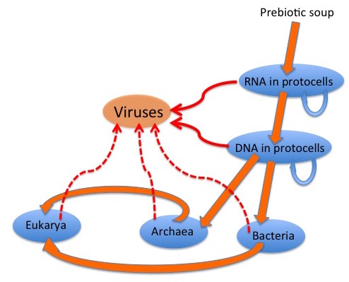
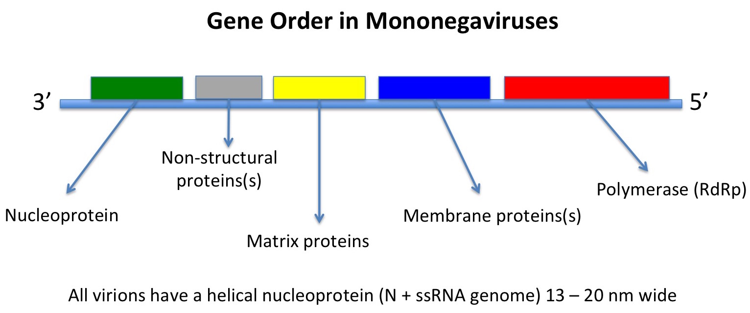
the viruses being very similar in genome organisation and indeed genome sequence.

SARSCoV isolates from humans bind angiotensin-converting enzyme 2 (ACE2) in order to gain entry into cells.  While the virus is very closely  related to isolates found in several Himalayan palm civets and a raccoon dog in Chinese live-meat markets, palm civets in particular outside markets were largely free from SARS-CoV infection – indicating to epidemiologists that this was not the natural animal reservoir of SARSCoV.  Other studies determined that there was a group of CoVs very similar to SARSCoV in horseshoe bats – but that these viruses differed from human isolates mainly in the N-terminal regions of their S proteins, which enable virus entry into host cells.  The SARS-like CoV (SL-CoV) S proteins have significant sequence divergence in the receptor-binding domain (RBD) from SARS-CoV S protein RBDs, including two deletions of 5 and 12 or 13 aa, and it has been predicted that SL-CoVs would not use ACE2 as a receptor.

In the absence of infectious isolates of SL-CoVs, these authors tested this hypothesis using an HIV-derived pseudovirus system: this used luciferase-expressing HIV-derived DNA constructs co-transfected into HeLa cells stably transduced with the ACE2 receptor gene from human, bat or civet, together with S gene-expressing plasmid constructs.  The system results in HIV-like virions containing RNA which expresses luciferase, with S protein on their surfaces.  Binding of pseudovirions to cells and their subsequent uptake was assayed by luminometry after addition of the luciferase substrate to cell lysates.

They found that that the bat SL-CoV S protein was unable to use ACE2 for cell entry regardless of the origin of the ACE2, and that the human SARS-CoV S could not use bat ACE2 as a functional receptor. Interestingly, after replacement of a small segment SLCoV S protein by the cognate sequence of SARSCoV S, the SL-CoV S protein also bound human ACE2.

The authors claim that this study reveals the “first example of host switching achievable for G2b CoVs [the taxonomic group including SL-CoVs] under laboratory conditions by the exchange of a relatively small sequence segment”.  They speculate that, given that bats may be coinfected by several CoVs, and that they associate at very high density, and CoVs have a tendency towards recombination, that it is reasonable to assume that bats act as a natural mixing vessel for CoVs, which can result in the emergence of novel viruses which could easily cross species barriers.

Adding fuel to the speculative fire is another paper in the same issue: this reports that there is evidence of a recombinant origin for SL-CoVs, and there is probably “…an uncharacterized SLCoV lineage that is phylogenetically closer to S[ARS]CoVs than any of the currently sampled bat SLCoVs.”

So let’s all just wait for the next one, shall we?