Archive for the ‘General Virology’ Category

DNA Preparation Tubes Contaminated with Novel ssDNA Virus

21 September, 2013

See on Scoop.itVirology News

A novel virus thought to have come from human samples appears to have been derived from seawater during the manufacture of tubes used to extract DNA.  

Ed Rybicki‘s insight:

I have a problem with the original report, in J Virol (http://jvi.asm.org/content/early/2013/09/05/JVI.02323-13.abstract?related-urls=yes&legid=jvi;JVI.02323-13v1): not that they discovered it, because that was done well.  However, they essentially REdiscovered something that was ±100% identical to a virus already sequenced and named by Chinese researchers – who did not use the Qiagen kits, apparently – and then gave it a new name!


Sorry, that is simply bad practice!  It also smacks of scientific imperialism of a sort that characterised early discovery work on HTLVs and on HIV, when US researchers calmly treated earlier characterisations as if they had never happened.


There is another leap that I do not think is justified: the authors claim that


"Analysis of environmental metagenome libraries detected PHV sequences in coastal marine waters of North America, suggesting that a potential association between PHV and diatoms (algae) that generate the silica matrix used in the spin columns may have resulted in inadvertent viral contamination during manufacture".

Really?  On the basis of presence of a sequence in a metagenomic trawl?  No resampling with specific primers on a fresh sample?   And surely the generation of the silica matrix is done under conditions that would totally destroy adventitiious DNA?

So – an interesting paper, and a valuable notification (although it might have been nicer if they’d shared their findings informally, to save other people like our Virology Diagnostics lab time and money).  But flawed, in my opinion.

See on www.the-scientist.com

Moratorium on using live rinderpest virus lifted for approved research

30 July, 2013

See on Scoop.itVirology News

Benefits of future research should be carefully balanced against potential risks

Paris, 10 July 2013 – A moratorium on using live rinderpest virus for approved research has been lifted by the Food and Agriculture Organization of the United Nations and the World Organisation for Animal Health (OIE).

The moratorium followed the adoption of a Resolution in May 2011 by all OIE Member Countries that urged members to forbid the manipulation of rinderpest virus containing material unless approved by the Veterinary Authority and by FAO and OIE.

The two organizations have now put in place strict criteria and procedures to follow in order to obtain official approval for any research proposals using rinderpest virus and rinderpest virus-containing materials. One of the most crucial requirements is that the research should have significant potential to improve food security by reducing the risk of a reoccurrence of the disease. This procedure replaces an earlier complete ban on handling the virus.

Rinderpest was formally declared eradicated in 2011, but stocks of rinderpest virus continue to exist in laboratories. In June 2012, a moratorium on handling the virus was imposed after an FAO-OIE survey found that the virus continues to be held in more than 40 laboratories worldwide, in some cases under inadequate levels of biosecurity and biosafety.

When rinderpest was officially eradicated, FAO and OIE member countries committed themselves to forbid the manipulation of rinderpest virus-containing material unless approved by the national veterinary authority as well as by FAO and OIE.

Paramyxovirus EM courtesy of Linda Stannard

Thanks to Len Bracher for alerting me to this.

Ed Rybicki‘s insight:

This is an interesting sequel to the eradication of wild rinderpest virus, which I have covered in some detail here on ViroBlogy: see here (https://rybicki.wordpress.com/2010/11/05/rinderpest-gone-but-not-forgotten-yet/) and here (https://rybicki.wordpress.com/2011/08/03/deliberate-extinction-now-for-number-3/).

The article covers an interesting prospect: that it may be possible to use attenuated, safe vaccines against the related peste des petits ruminants virus (PPRV) not only to protect against any resurgence of rinderpest, but also to eradicate this rather nasty virus.

Which is, apparently, spreading at rather an alarming rate, and is an obstacle to small ruminant production (http://www.fao.org/ag/againfo/resources/documents/AH/PPR_flyer.pdf).

So maybe this is “Now for Number 4!” time.

See on www.oie.int

Maize streak virus revisited: 25 years on

20 March, 2013
Maize streak virus: photo from 1978

Maize streak virus: photo by Robert G Milne in Cape Town from 1978

Twenty-five years ago, I wrote a brash, naïve little piece entitled “Maize streak virus virus: an African pathogen come home?” for the South African Journal of Science, laying claim to a virus that we had just started working on – Maize streak virus (MSV) – on the basis that it had first been described from this country in 1901, that it was endemic here, and that it still caused major crop losses.  I did this because research on this and related viruses seemed to have moved almost completely offshore, to Europe and the USA, and

“…the most interesting of the viruses that grow all around us have already been whisked away to foreign laboratories; [that] there they have been cloned, sequenced, and had their most intimate details exposed, far from their native shores”. [Yes, I really did write like that back then].

I asked at that time, if we should

“…perhaps be content to supply foreigners with the (pathogenic) fruits of our fields, and to marvel when the answers come filtering back from abroad?”.

I answered myself by saying that

“…prospects for worthwhile research on African geminiviruses, and on any other indigenous pathogens, are at least as good here as anywhere else.  Our facilities are the equal of those abroad, the necessary expertise is certainly not lacking, and the viruses are on our doorstep.”

I’m a little shocked now that I could have said that then: the paper quotes only three pieces of work from our lab, one of them a Masters dissertation and two papers done by my erstwhile supervisors; we had not yet sequenced any virus, let alone a geminivirus, and all we had was brashness and hope.  Indeed, I went on to say the following:

“We are, incidentally, the only research group with access to molecular biological techniques which is actually working on the virus in its natural environment: this is very useful, as with the virus in all its forms and its vector(s) literally on our doorstep, we can rapidly accumulate, identify and characterize distinct isolates for study here or elsewhere.  We hope there will be a little more of the ‘here’, and a little less of the ‘elsewhere’, from now on”.

I outlined what it was that we ambitiously wanted to do – seeing as we had no money, and only one PhD student at the time – as follows:

“…we now have distinctly different genomic maps of three isolates [!] which differ in serology and symptom expression; we have cloned genomic DNA of several more isolates, and can potentially clone and [restriction] map many more.  With this type of work now solidly established, we intend to investigate other biological variants of MSV – and other native cereal geminiviruses – in maize, cereal grains and other members of the Gramineae.  The aim is to explore the genetic diversity of naturally occurring types of MSV and related viruses, and to identify any isolates that appear unusual in terms of symptom expression, serology or transmission.  These would be interesting to map, and potentially useful in recombinational analyses for the fine mapping of determinants of pathogenicity and host range.” [see later]

The article obviously sank without trace: I can find only three citations to it; two of them mine, and the third from a South African maize breeder.  How the overseas labs that I compared us to must have sniggered…actually, I doubt that happened at all; I am sure none of them ever read it!  In retrospect, we really were regarded as a backwater, and as wannabe geminivirologists; I had at least one collaboration request rebuffed with “we don’t feel our work would be advanced by working with you”, and was told “we’re already working on that, so you shouldn’t bother” for a couple of other proposals.

My hubris was not entirely misplaced, however: we did in fact go on to develop into a world-leading MSV and geminivirus molecular virology laboratory; it just took another fifteen years or so!

So where are we, twenty-five years on from my cheeky article?  Much water has flowed under several bridges; I expanded from molecular virology in the 1990s into plant and vaccine biotechnology in the 2000s, while keeping a geminivirus research group going – and we have published and co-published something like 55 peer-reviewed journal articles and several encyclopaedia and book chapters on MSV and other “African streak viruses” alone, let alone another 14 or so articles on other geminiviruses, with some 1200 citations.  We have papers on geminivirus mapping and sequencing, virus diversity, biogeographical variation, quantitation of symptoms, molecular determinants of pathogenicity, recombination, engineering maize for resistance, the use of two of the viruses as gene expression vectors – and cover pictures for Plant Biotechnology Journal and Journal of Virology.

Cover Illustration: J Virol, October 2011, volume 85, issue 20

Cover Illustration: J Virol, October 2011, volume 85, issue 20

I started with one Honours student in 1986, who went on to do a Masters in 1988; we moved on to having one PhD student in the late 1980s to up four PhD students simultaneously in the mid- to late 1990s, and a postdoc at the same time.  The projects went from simple diversity studies of a few viruses using restriction mapping, through the application of PCR, to partial genome sequencing and studying the molecular biology of infectious clones of the viruses, with a very profitable sideline in phylogenetic analyses; we also moved – with Professor Jennifer Thomson – into a parallel track of plant biotechnology, aimed at engineering resistance to MSV in maize.  We added another track early this century, working on similar ssDNA circoviruses of parrots, using all of the expertise we had accumulated on geminiviruses.  We truly work on “circomics” now – the study of small circular genomes – with its subsets “geminiviromics” and “circoviromics”, with a library of literally hundreds of sequenced MSVs and distinct grass mastreviruses and BFDVs.

Geminivirus particle: characteristic doubled icosahedron containing a single ssDNA

Geminivirus particle: courtesy of Russell Kightley Media

The geminiviromics group has pretty much got away from me now; the folk I trained as PhD students in the late 1990s and early 2000s were enthused enough with the field that they have gradually usurped my leadership and supervisory role, and made the field their own.  I still maintain an interest in using Bean yellow dwarf mastrevirus (BeYDV) as an expression vector for “biofarming” purposes; I am also maintaining a project on Beak and feather disease circovirus (BFDV) diversity and plant-made vaccines.  I think we pretty much did what we set out to do – including the brave prediction I made about host range and pathogenicity, which led to some very interesting work on recombination and genome modularity, and the successful engineering of pathogen-derived resistance to MSV.

So I owe some thanks, in retrospect: first, to Barbara von Wechmar, who sparked the interest – and provided isolates, leafhoppers, and expertise.  Second, to Bev Clarke and Fiona Tanzer (aka Hughes), who were brave enough to blaze the trail, and clone our first MSVs – and make one infectious, in the case of Fiona.  Thanks to Wendelin “Popeye” Schnippenkoetter, for your single-minded perseverance in mixing and matching genomes; thanks Kenneth Palmer, for showing the way for transient expression assays in maize cells and engineering MSV as a vector.  Thanks Janet Willment, for mapping replication origins in MSV and expanding us into wheat viruses; thanks Jennifer Thomson for the collaboration, and Fiona and Tichaona Mangwende and Dionne Shepherd for breaking us into maize resistance engineering.  Thanks Christine Rey for the collaboration, and Leigh Berrie for your quiet competence in our detour into South African cassava mosaic virus.  Thanks Darrin (aka Darren) Martin and Eric van der Walt, for so brilliantly exploring MSV diversity, evolution and recombination – and Darrin for endless amusement in the lab, as well as for two completely distinct and invaluable software packages, for symptom quantitation and recombination analysis.  In the present generation, thanks to Suhail Rafudeen and our student Rizwan Syed (and Dionne and Darrin as supernumerary supervisors); thanks Aderito Monjane for doing such a ridiculous amount of work for a superlative PhD; thanks Dionne and Marian, for keeping the maize engineering afloat – and thanks also to Arvind Varsani, for retraining himself from a papillomavaccinologist to a circomicist, and for popping up everywhere.

Vaccines: a simple message

28 February, 2013

+MaryMangan over there on Google+ made an interesting point about simple messages to refute the kinds of nonsense promulgated by vaccine denialists, among others.

Here’s my contribution:

Vaccines!

Vaccines!

TMV in mouse lungs: more thoughts and refutations

13 February, 2013

tmv sedimhave been thinking about this paper (see last post), and it and other people’s posts (eg: Tommy Leung’s) have prompted more response.

I note the authors  say the following:

“There is other published literature that challenges the dogma of the strict boundaries between plants and vertebrates for viruses. In non-vertebrate animals, it was shown that plant pathogenic viruses displayed complex interactions with insects, and the transcription and replication of some plant viruses within insects was described [29][32]. In addition, in some cases, insects were found to be affected by plant viruses [33]. Furthermore, it was recently shown that Tomato spotted wilt virus (TSWV) could infect two human cell lines, HeLa and diploid fibroblasts, depending on the expression of a viral polymerase-bound host factor[34]. Additionally, despite plant virus replication was not observed in animals, Cowpea mosaic virus (CPMV), a plant comovirus in the picornavirus superfamily, was able to bind and enter mammalian cells, including endothelial cells, and the binding protein for the virus was identified as a cell-surface form of the intermediate filament vimentin [35]. Furthermore, CPMV was found to persist for several days post oral or intravenous inoculation in a wide panel of body tissues in mice, including in the lung and the liver [36]. Additionally, it was demonstrated that TSWV induced a strong immune response in its insect vector Frankliniella occidentalis [37] and that oral administration of Cowpea severe mosaic virus, Alfalfa mosaic virus and chimeric plant virus particles induced a durable and systemic immune response in mice [38][39]

Yes.  Um. Well.  The “dogma of the strict boundaries between plants and vertebrates for viruses”?  I have been teaching virology for 32 years, and I am not aware of actual DOGMA – as in, “that which has to be believed”.  Rather, there has been the cumulative set of OBSERVATIONS that nothing that anyone has ever isolated out of a plant – and that replicates in it – has infected a vertebrate.  I make that distinction, because there is always the possibility that, as we and others have found with insect viruses, plants can act as a “circulative, non-propagative vector” for insect viruses (for Rhopalosiphum padi aphid virus in barley, from my lab, and Leafhopper A virus in maize) – and if one realises that male mosquitoes, and often also females, feed on plants…you see where I’m going here?  As in, it might well be possible for a virus that multiplies in an insect and also in a vertebrate, to POTENTIALLY be found in a  plant?

In ay case, this is largely beside the point, because the authors get sidetracked into discussing Tomato spotted wilt – which happens to be a plant-adapted bunyavirus, most closely related to insect and vertebrate phleboviruses – “depending on the expression of a viral polymerase-bound host factor”.  Really??  And if it isn’t there?  Does the virus in fact spread?  For that matter, my lab has cell-free translated two aphid picorna-like virus genomes in rabbit reticulocyte lysates, but we made no claim that it could happen in rabbit cells.  Moreover, they make much of the fact that “a plant comovirus in the picornavirus superfamily, was able to bind and enter mammalian cells…[and] was found to persist for several days post oral or intravenous inoculation in a wide panel of body tissues in mice, including in the lung and the liver”.

Yes?  And?  A REALLY stable plant virus was able to bind and enter animal cells, and persist?  The problem with that is…?

We in the virus-like particle vaccine field RELY on the fact that VLPs will be taken up by cells of the immune system in vertebrates, and that they will elicit immune responses – so why is this regarded as a problem?  In fact, TMV has itself been tested as an RNA vaccine delivery system, due to its ability to protect a RNA payload, and get itself delivered into reticulocytes and macrophages – meaning this property has been known for some time, and has not hitherto been seen as a problem!

I think these authors have hyped something that is quite interesting into what THEY regard as a potential problem, for the purposes of getting their article accepted – and I think this needs to be recognised, and that the perceived risks need to be minimised by the knowledgeable.

PLOS ONE: Tobacco Mosaic Virus in the Lungs of Mice following Intra-Tracheal Inoculation

13 February, 2013

See on Scoop.itVirology News

“Plant viruses are generally considered incapable of infecting vertebrates. Accordingly, they are not considered harmful for humans. However, a few studies questioned the certainty of this paradigm. Tobacco mosaic virus (TMV) RNA has been detected in human samples and TMV RNA translation has been described in animal cells. We sought to determine if TMV is detectable, persists, and remains viable in the lung tissues of mice following intratracheal inoculation, and we attempted to inoculate mouse macrophages with TMV. In the animal model, mice were intratracheally inoculated with 1011 viral particles and were sacrificed at different time points. The virus was detected in the mouse lungs using immunohistochemistry, electron microscopy, real-time RT-PCR and sequencing, and its viability was studied with an infectivity assay on plants. In the cellular model, the culture medium of murine bone marrow derived macrophages (BMDM) was inoculated with different concentrations of TMV, and the virus was detected with real-time RT-PCR and immunofluorescence. In addition, anti-TMV antibodies were detected in mouse sera with ELISA. We showed that infectious TMV could enter and persist in mouse lungs via the intratracheal route. Over 14 days, the TMV RNA level decreased by 5 log10 copies/ml in the mouse lungs and by 3.5 log10 in macrophages recovered from bronchoalveolar lavage. TMV was localized to lung tissue, and its infectivity was observed on plants until 3 days after inoculation. In addition, anti-TMV antibody seroconversions were observed in the sera from mice 7 days after inoculation. In the cellular model, we observed that TMV persisted over 15 days after inoculation and it was visualized in the cytoplasm of the BMDM. This work shows that a plant virus, Tobacco mosaic virus, could persist and enter in cells in mammals, which raises questions about the potential interactions between TMV and human hosts.”

Ed Rybicki‘s insight:

Interesting paper!  Which proves…which proves…which proves TMV is seriously resistant to degradation in animals and in mammalian cells; that it can enter macrophages; and that it…what?  What, exactly, are the “…questions about the possible interactions…”?  What would TMV do in mammalian cells?  Yes, it might be uncoated and be translated; it is far less likely that it MIGHT be able to replicate its RNA – and then?  While it can apparently be taken up quite efficiently by macrophages – a property which, incidentally, has led to its being trialled as an RNA vaccine delivery system – this is a dead end, and one that is quite normal for particles of any kind being introduced into mammals.

Which is something that happens every day, as we and our cousin mammals eat: it has been shown elsewhere that animals are actually quite good spreaders of plant viruses, some of which – like TMV and the even tougher Cauliflower mosaic virus – pass right through at high survival rates, and remain infectious.  We will all probably have eaten many grams of various viruses in our lives, and derived nothing more than nutrition from them.

I also remember, even though it was very late at night, 31 years ago, and in a bar in Banff in Canada, a conversation with one Richard Zeyen.  He told me they had used ELISA to test everyone in their lab for antibodies for TMV, seeing as they worked with it, and had newly developed a test.  And everyone was immune – presumably, to aerosolised TMV that had been breathed in or otherwise ingested.  Proving…that oral vaccines based on TMV could work, and that most of us are probably immune to all sorts of viruses that don’t replicate in us – and nothing more!

Including, in the case of many people in the Eastern Cape Province of South Africa, sampled by one Don Hendry via the local blood bank, to a virus of Pine Emperor moths – because it multiples to such high levels in its host that anyone walking in the pine forests was bound to be exposed via the environment.

So this is an interesting paper – and no more.  It will, of course, lead to alarmist articles and blog posts, and people calling out for urgent surveillance of food, in which people will find many viruses.  And so what?  They have been with us for as long as we have been eating plant-derived food, and have NEVER been associated with any disease, transmissible or otherwise – so my best advice is that we ignore them.

See on www.plosone.org

ViroBlogy: 2012 in review

1 February, 2013

So: thank you, anyone who clicked in, and regular visitors.  You make it worthwhile!!

The WordPress.com stats helper monkeys prepared a 2012 annual report for this blog.

Here’s an excerpt:

4,329 films were submitted to the 2012 Cannes Film Festival. This blog had 33,000 views in 2012. If each view were a film, this blog would power 8 Film Festivals

Click here to see the complete report.

CCHFV in South Africa

1 February, 2013

I am indebted to the National Institute for Communicable Diseases (NICD) in Johannesburg for their very informative newsletter, from which I culled this.

I would also like to very sincerely congratulate Professor Barry Schoub, a long-time former Director of the NICD, on his  African Society for Laboratory Medicine (ASLM) Lifetime Achievement Award!  Very well deserved.

Crimean-Congo haemorrhagic fever

Two cases of Crimean-Congo haemorrhagic fever (CCHF)  acquired in South Africa have been laboratory confirmed  in January 2013.

On 1 January 2013, a 31-year-old male working as a  game warden on private game ranch near Jagersfontein  (Free State Province) presented with clinical features  suggestive of CCHF. The patient did not report any tick  bites or direct exposure to unprocessed meat or  slaughtering of animals. The Centre for Emerging and  Zoonotic Diseases of the NICD/NHLS confirmed infection  with CCHF virus by PCR and serology testing.

A second case of CCHF was laboratory confirmed on 12  January 2013 in a 44-year-old male hospitalised in  Bloemfontein, Free State Province. He had been on a  farm in Pomfret, North West Province (situated ±5 km  from the border with Botswana), where he was bitten by  a tick. Three days later he developed symptoms, and  presented with fever, rash, conjunctivitis and pharyngitis.  No laboratory-confirmed cases were identified in 2011- 2012.

Human CCHF cases have been reported annually  from South Africa since 1981, when it was first  recognised in the country; between 0 and 20 cases of  CCHF are diagnosed each year. Through nearly thirty  years of passive surveillance, a total of 187 cases has  been laboratory confirmed. Although cases have been  reported from all of the nine provinces, more than half of  the cases originate from the semi-arid areas of Northern  Cape Province (31.5% of cases) and Free State Province  (23% of cases).

CCHF infection is generally asymptomatic in many species  of wildlife (including antelope) and livestock animals  (including cattle, sheep, goats, hares and ostriches).  Humans  become  infected  sporadically  by  ticks,  particularly  Hyalomma ticks, which are both reservoirs  and vectors for CCHF virus. Other modes of transmission  include direct contact with blood/tissues of infected  animals, and in the case of healthcare workers, through  direct contact with the blood/tissue of infected patients;  nosocomial outbreaks are well described and have been  associated with high mortality rates. Disease may be  severe in people, with case-fatality rates reported as 3 –  30% across various studies.

Detailed information for healthcare workers regarding  CCHF can be found on the NICD website  http:// http://www.nicd.ac.za/ (see General Public FAQ, or Health Workers FAQs here).

Virus-like particle and Nano-particle vaccines 2012: a conference report

30 January, 2013

Alta van Zyl, Virology Group, Molecular & Cell Biology Department, UCT

Introduction:

VLP flusm

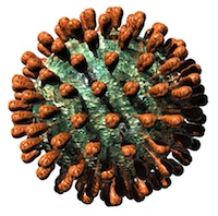
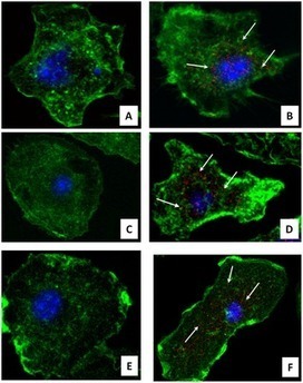
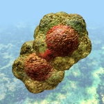
Haemagglutinin-only Influenza A virus VLP. Courtesy of Russell Kightley Media

The new international conference on virus-like particles and nano-particles (VLPNPV) took place in Cannes, France at The Novotel Montfleury Hotel from the 28th to the 30th of November 2012.  The scope of the conference included virus-like particles (VLPs), the plant-based expression of VLP vaccines as well as expression and optimisation of VLPs.

Other topics included in the conference were:

  • VLP platform delivery systems
  • VLP vaccines
  • Nano-particles and nano-particulate vaccines

A multitude of topics were covered during the conference and many of the talks pertained to the immunogenicity of the VLPs and nano-particles and how they compared with the immunogenicity of DNA or subunit vaccines.

Talks were given by researchers from companies such as Medicago, Mucosis, Pevion Vaccines and Novavax. These talks gave a perspective on factors that need to be considered when commercialising VLP/nano-particle vaccines and to be GMP compliant.

Compelling presentations:

Developing plant-made virus-like particle vaccine products: An integrated platform from discovery to commercial scale

Marc-Andre D’Aoust, Nathalie Landry, Sonia Trepanier, Michele Dargis, Manon Couture and Louis-Philippe Vezina (Medicago, Quebec City, Quebec, Canada)

This talk was about a plant-made VLP against both pandemic and seasonal influenza- these vaccines are now in the clinical trial phase. What was especially interesting was the view from an industry point of view where expression had to be scaled up to produce large amounts of vaccine.  The Medicago platform can synthesize and clone approximately 100 gene constructs in two weeks, they can prepare 100 bacterial cultures per week and they have automated infiltration where 200 plant transformations can be performed per day and 150 VLP engineering approaches can be tested in one week.  For influenza Medicago tested 48 different infiltration approaches in one day for HA, NA, M1, M2 as well as P1 Gag and HGalT.  Medicago has been able to produce 10 million doses of HA VLPs in just one month.

See also: 

  • D’Aoust et al (2010) PBJ 8:  607-619 – The production of hemagglutinin-based virus-like particles in plants: a rapid, efficient and safe response to pandemic influenza.
  • http://www.medicago.com

Development of RNA-free plant VLPs a source of novel therapeutics

George Lomonossoff (John Innes Centre, Norwich, UK)

This group made empty Cowpea Mosaic Virus (CPMV) VLPs that contained no RNA.  CPMV VLPs are versatile nanoparticles to which organic, inorganic and biological molecules can be bound.  The empty nature of the particle means that they can be used as carrier molecules for therapies; this could prove to be potentially useful as a cancer-treatment therapy.  The system is advantageous because of the lack of RNA which makes the particles non-infectious and no bio-containment is needed for the production of these VLPs.

Immunogenicity of VLPs: an immunological perspective

Martin Bachmann (University of Zurich, Zurich, Switzerland)

Background was given from immunological point of view about what makes VLPs so immunogenic. Three properties contribute to the immunological properties of VLPs (1) their size, (2) the repetitiveness of the particle capsid which provides multiple sites for antibody binding and (3) TLR ligands – the particle can be disassembled, the RNA removed and replaced with a TLR ligand to enhance immunogenicity. Also, the size of VLPs is optimal for drainage to the lymph nodes.

Immunogenicity optimization strategies for public-sector development of vaccines: the critical role of optimizing the antigen.

Martin Howell Friede (WHO, Geneva, Switzerland)

This talk was about looking at VLPs from the vaccine development view.  Monomeric antigens are not very immunogenic; therefore adjuvants were developed and came into use. For an efficient vaccine the antigen must be multimeric as antigen alone is insufficient to be immunogenic without adjuvant. Two factors have to be considered when producing a vaccine for FDA approval; (1) optimise the antigen before using an adjuvant, (2) use an adjuvant that has already been approved by the FDA. VLPs as vaccines provide the potential for immune-stimulation without the addition of adjuvant as the multimeric presentation of the antigen will enhance its immunogenicity.

Enhancing the immunogenicity of VLP vaccines

Richard W. Compans (Emory University, Atlanta, Georgia, USA)

This talk highlighted strategies which could be used to enhance the immunogenicity of VLPs.

  1. Look at alternate routes for vaccine delivery (intranasal, intramuscular, subcutaneous etc)
  2. Increase the breadth of immunity by enhancing responses to conserved antigens/epitopes
  3. Increase the amount of antigen incorporated into VLPs
  4. Incorporate the adjuvant into the VLPs as part of the structure

See also:

  • Ye et al (2011) PLoS One 6(5):  e14813
  • Wang et al (2008) J Virol

Innate and adaptive responses to plant-made VLP vaccines

Brian Ward (McGill University, Montreal, Quebec, Canada)

Brain Ward is also the medical officer at Medicago.  Humans rarely react to plant proteins/antigens. The plant glycans fucose/xylose at the N-terminal is an allergen and can cause anaphylaxis in humans. During trial experiments with influenza no individuals developed IgE responses to plant glycans, therefore plant produced vaccine is safe. The H1 VLP induced long lasting memory multifunctional T-cell responses in humans.

Impressions of the conference:

The conference was well organised with leaders in the field presenting their work. Interaction with the delegates aid in building crucial networking opportunities and work relationships. The international arena is packed with new technology development allowing us the opportunity to learn and improve our own understanding of various concepts.

This conference proved to be an invaluable learning experience and I thank the NRF for this opportunity and for providing me with the funding to attend this conference.  The exposure to conferences, especially those in the international arena, aid in the development of students and contribute to the quality of research that is conducted at UCT.

References:

1. VLPNPV website

(http://www.meetingsmanagement.co.uk/index.php?option=com_content&view=article&id=33&Itemid=83)

2.  Personal notes taken at the conference

And so it went – 2012, that is

14 December, 2012

…like a rocket…flashed past; I’m still emotionally in August or so!

I meant to do some more substantive posts instead of only copying Scoop.it Virology News posts here; however, the best-laid plans and such, and I didn’t.  I will in 2013, though – and there will be an iBook coming or possibly even two (influenza and PCR), so I will use this forum to announce glad tidings.

Then there’s the ZA Virus [=Zombie Apocalypse, obviously] novel, and Green Vaccines, and…OK, getting ahead of myself here!

Thanks for the support and readership, I hope everyone has a good solstice break!

Best,

Ed

PS: some access stats for 2012 for you.  Looks like the only places that DON’T access ViroBlogy are parts of central and west Africa, central Asia and Greenland.

Screenshot 14:12:2012 12:11

Screenshot 14:12:2012 12:09


Screenshot 14:12:2012 12:20