Posts Tagged ‘Evolution’

What are you looking for?

18 April, 2011

It is quite educational sometimes, to look and see what it is people are looking for, when they end up on ViroBlogy.  Here are the queries over the course of the last month:

Search Views
ebola 577
ebola virus 64
rolling circle 44
how did viruses evolve 29
mimivirus 21
despair posters 19
flaviviridae 17
lassa fever 14
bacterial dna replication 11
ebola virus patients 11
viroblogy 9
where did viruses evolve from 9
ebola pictures 9
rift valley fever 9
what did viruses evolve from 9
geminivirus 8
west nile virus structure 8
ebola patient 8
where h1n1 came from 7
ebola virus pictures 7

I count 6 mentions of Ebola, 4 of virus evolution generally…and a gratifyingly high number of queries on rolling circle replication, given our lab’s interest in it.  Of course, I have had long had an obsession with Ebola and similar nasties, and have been deeply interested in the evolution of viruses ever since I started studying them, so it is good to see others share my interests!

So in the spirit of giving people what they want, look for blog posts with titles like “The evolution of viruses using rolling circle replication: its complete non-relevance to Ebola and H1N1 influenza viruses”.  But seriously – I will be putting up some more posts around the most popular subject areas, if only to help make sure that The Truth Gets Out There.

Ed Rybicki

Viruses as nanomachines! Or: what you can believe from YouTube

6 December, 2010

I have for some years now been teaching my undergrad students that virus particles are nanomachines: that is, they are highly sophisticated nanoscale (read: ultramicroscopic) devices whose function is to specifically deliver genetic material into an environment where it can be expressed and replicated, so as to make more virus particles.

Nanoscale von Neumann machines, then – and if you want to see what a macroscale vN machine could do, just watch “2010: Odyssey Two“.

Ah, but what’s a von Neumann machine, you ask?  Well, I note Wikipedia has the following:

  • Self-replicating machines, a class of machines that can replicate themselves
  • Universal Constructors, self-replicating cellular automata
  • Von Neumann probes, hypothetical space probes capable of self-replication
  • Nanorobots capable of self-replication

I especially like the last two – because, as I showed in a previous blog post, I like the idea of virus particles or virions as “inner space craft”.  That this neatly marries my recreational and professional reading is no coincidence – because they cross-pollinate one another, in that I get ideas about the nature of viruses from SF, and my virology training informs scenarios I would like to write about.  Someday.  Soon, possibly.  Really.  Instead of writing about parallel universes contactable via the internet….

However, there is more to viruses and nanotechnology than phages with contractile tails, whether or not they have been around for billions of years: mimiviruses too have both nanoscale DNA loading and rapid-delivery systems, as previously discussed here.

Although I have a passing fondness for possibly my most successful animation – made from actual EMs, done by Linda Stannard.

T4 phage infecting a cell

So it was with some pleasure I saw recently on YouTube a video labelled “Viruses are nanotechnology (how a virus works)“.  I was a little less pleased when a voice confidently announced that “…a virus isn’t alive, people – it’s non-metabolising…”, as if that was the sole and necessary criterion for life.  I am at one with another Polish-named person – one Bernard Korzeniewski – in thinking  that life is (from MicrobiologyBytes)

The phenomenon associated with the replication of self-coding informational systems” © E.P. Rybicki, 1996. Incidentally, I find another person with a Polish name has said something very similar, in 2001 – which means it must be true. Bernard Korzeniewski describes life as: “A network of inferior negative feedbacks subordinated to a superior positive feedback.”

See, no mention of metabolism – or even of cells!  But what got the hairs on the back of my neck standing up, however, was some of the rest of it – delivered in a smooth, folksy manner, with stunning video footage.  Absolute cr@p, most of it: viruses are too complicated to have evolved, so they have to be alien nanotech???

Obviously some weird kind of conspiracy theory cross technobabble – but very seductive, to the uninformed.  Some of the comments are also just out of this world – literally!

Fortunately, there are some real science videos out there too – some of which I have also used in lecturing, if only to illustrate just how cool structural biology can be when used to study viruses.  Prime among these is one of T4 virus (Enterobacteria phage T4) infecting E coli; another magical one  from the same source is a depiction of the molecular motor which winds DNA into T4 heads.  A longer video has Michael Rossman, whose lab did the structural work behind the videos, explaining how the phenomenon could be useful in understanding viruses like herpesviruses in humans, which also appear to have molecular motors for DNA delivery – and, of course, how we can mess with them.

Self-assembly of viruses is also a good topic for video – and the full-length  Seyet T4 video is stunning in this regard.  So too is this one, showing a PhiX174 microvirus particle assembling.  One of my favourites, though, is the simplest: this is the depiction of how simple shapes can be induced to self-assemble into a virus-like particle – just by shaking.

I suppose, like everything, you get what you pay for with YouTube: which is nothing, most of the time.

But every now and then, a gem – which is what makes it fun to look.  I’m off to hunt down a Rolling Stones video virus replication videos!

The largest marine virus yet

13 November, 2010

This is another welcome guest post from Gillian de Villiers, a Scientific Officer in our Vaccine Group.  This was presented as a Journal Club article recently, and fit so well into my continuing theme of “viral diversity from water” that I asked her to write it up.  Thanks Gillian!

Giant virus with a remarkable complement of genes infects marine zooplankton

Matthias G. Fischer, Michael J. Allen, William H. Wilson, and Curtis A. Suttle

PNAS published ahead of print October 25 2010 www.pnas.org/content/early/2010/10/15/1007615107

This publication covers the sequencing of the genome of Cafeteria roenbergensis virus(CroV).  This nucleocytoplasmic large DNA virus (NCLDV) is the largest marine virus described to date, and its closest relative is Acanthamoeba polyphaga Mimivirus.

Among the questions raised in this paper are:

  • what is the evolutionary origin of big viruses?
  • Did they get their genes from horizontal gene transfer (including from eukaryotes), or
  • are the “eukaryotic” genes viral in origin?

Spoiler alert: the authors do not answer this question.

Please note: this is a virus from a seawater host.  It is the largest marine virus yet found, but how hard has anyone been looking?  This ties in with Ed’s theme that we should be looking for viral diversity and interesting things in the water, because interesting things have been found there.

Some background…

This lytic virus strain was isolated off the coast of Texas in the 1990s.  The host, Cafeteria roenbergensis was originally misidentified as a Bodo species.  It is a major micro flagellate grazer (microzooplanton = major ocean predator) a 2-6um “bicoecid heterokant phagotrophic flagellate” and has been found in multiple marine environments including surface waters, deep sea sediment and hydrothermal vents.

In other words, the host is an extremely significant part of the ocean ecosystem, and has been found in most places.  The authors note that protists host the largest viruses known and that other giant viruses probably are widespread in the oceans, but so far only the Acanthamoeba-infecting giant viruses have been characterised (Acanthamoeba does not live in the ocean). Viral infections of cyanobacteria play a significant role in global oxygen production; in a similar way the viral infections by CroV may have implications for carbon and other nutrient cycling and the “food chain” in the oceans, although this is beyond the scope of the article.]

Results

The genome is the second-largest viral genome described and at 730kb is very AT rich.  Approximately 618kb is thought to be coding with 544 predicted protein-coding genes.  At least 274 genes are expressed during infection.  22 percent of CroV CDSs (coding sequences) were probably best related to eukaryotic genes.  Most CroV CDSs had unknown function, but 32% of CDSs could be assigned a putative function.

For enzymatic functions that have not previously been reported in any other viruses you can refer to Table S1 of the Supplemental materials.

This is similar to CroV’s closest known relative, Mimivirus, where of 911 predicted genes only 300 were assigned a predicted function (see table).  Only 1/3 of their genes are common to these two viruses!  This suggests tremendous diversity within the nucleocytoplasmic large DNA viruses, as they may have common evolutionary origins for some genes, but not for others.  As viruses are not monophyletic (although the NCLDVs may be) and can be considered to be bags of protein that contain genetic material and share a strategy (rather than an origin) this may not be particularly surprising.  But I find it amazing that so many potential genes, and so many unique potential genes, have been found in these organisms.

Included in the genes assigned function are genes involved in translation.  CroV encodes an isoleucyl-tRNA synthetase and putative homologs of eukaryotic translation initiation factors.  22 tRNA genes and two putative tRNA-modifying enzymes: tRNA pseudouridine 5S synthase and tRNAIle lysidine synthetase were found.  Mimivirus also has four tRNA synthetases and several putative translation factors.

Cafeteria roenbergensis virus Acanthamoeba polyphaga Mimivirus
~730kb dsDNA genome ~1200kb dsDNA genome
300nm capsid 500-750nm capsid (publications differ)
Largest marine virus yet described Largest virus yet described
Second-largest virus yet described
544 predicted genes 911 predicted genes
174 genes with predicted function 300 genes with predicted function
Host: Cafeteria roenbergensis Host: Acanthamoeba castellani (amoeba)
Habitat: marine environment Habitat: soil (?freshwater)
Genes shared with Mimivirus ~ 1/3 Genes shared with CroV ~ 1/5

Similarly to other large DNA viruses a number of DNA repair genes were found.  This includes a base excision repair pathway that appears complete.  In addition crov115’s gene product is predicted to be a CPD class 1 photolyase, the first viral homologue in its class.  Crov149 appears to be part of a recently described photolyase/cytochrome group found in several bacterial phyla and euryarchaeotes, but not among established types of photolyase.  The authors suggest that the only eukaryote with this gene, Paramecium tetraurelia may have acquired it by horizontal gene transfer from a giant virus

CroV also has transcription-related genes including eight DNA-dependent RNA polymerase II subunits, six transcription factors involved in transcription initiation, elongation, and termination, a tri-functional mRNA capping enzyme, a poly (A) polymerase, as well as helicases.  Mimivirus provides considerably more genes for protein transcription and translation than most viruses, and sets up its own ‘virus factory’ in the cytoplasm of the cell.  It is possible that CroV has a similar strategy, with viral gene transcription independent of the host and occurring in the cytoplasm.

Of the three DNA topoisomerases, two are very similar to the counterparts in Mimivirus.  CroV TopoIB is the first viral homolog of the eukaryotic subfamily, but the Mimivirus TopoIB appears to be from the bacterial group.  Although the evolutionary origin appears to differ, the topoisomerases are presumably important in transcription, translation or packaging of giant virus genomes, as they appear in both CroV and Mimivirus genomes.

CroV has four inteins: self-splicing proteins.  They are found in DNA-dependent DNA polymerase B (PolB), TopoIIA, DNA-dependent RNA polymerase II subunit 2 (RPB2) and the large subunit of ribonucleotide reductase (RNR).  Inteins have previously been found in viruses infecting eukaryotes, including Mimivirus PolB.  CroV TopoIIA intein is the first case of an intein in a DNA topoisomerase gene.

Microarray analysis on the 12-18 hr infection cycle showed around half the predicted genes, and 63% of the tested genes were expressed during infection.  Work on Mimivirus and PBSC-1 showed transcription of nearly all predicted genes, so this work may underestimate the true transcriptional activity of CroV.  CroV gene expression has an early phase 0-3 hrs after infection affecting 150 genes, and a late phase affecting 124 genes 6 hrs or later post-infection including all the structural components predicted.  A conserved early promoter motif “AAAAATTGA” was identified in 35% of CDSs and is nearly identical to the Mimivirus early promoter motif “AAAATTGA”.  A promoter element for genes transcribed during the late phase of CroV infection was found that is unrelated to the putative late promoter motif in Mimivirus.

A genomic fragment involved in carbohydrate metabolism was also found.  This 38kb fragment includes enzymes for biosynthesis of 3-deoxy-D-manno-octulosonate (KDO).  This is part of the lipopolysaccharide layer in gram-negative bacteria and is found in the green alga Chlorella and the cell wall of higher plants. Ten of the enzymes involved in carbohydrate metabolism were expressed, suggesting a role in viral glycoprotein biosynthesis, suggesting the virion surface may be coated with KDO- or sialic acid-like glycoconjugates. 

There are no homologs in Mimivirus suggesting this region must have been acquired after the CroV and Mimivirus lineages split (or that the Mimivirus lineage lost it subsequently?).  This may have been acquired from bacteria, however GC content is even lower than for the rest of the CroV genome, and a number of the proteins are phylogenetically between bacterial and eukaryotic homologs.

Phylogenetics and Speculations

Phylogenetic reconstruction of NCLDV members. Redrawn and simplified from Fig. 4. The unrooted Bayesian Inference tree was generated from a 263-aa alignment of conserved regions of DNA polymerase B

CroV is an addition to the group of NCLDVs including Ascoviridae, Asfarviridae, Iridoviridae, Mimiviridae, Phycodnaviridae, Poxviridae and Marseillevirus, which are presumed to be monophyletic. CroV seems to be the closest known relative to Mimivirus although it is substantially smaller.  The topology of the NCLDV tree strongly suggests the five largest viral genomes (all mimiviruses) are more closely related to each other than to other NCLDV families.  They may have originated from an ancestral virus that was already an NCLDV that encoded more than 150 proteins.

Mimivirus is the most studied NCLDV, and is the largest.  Most Mimivirus genes have no cellular homologs and may be very ancient, with 1/3 of genes having originated through gene and genome duplication and less than 15% of the genes having potentially been acquired by horizontal gene transfer from eukaryotes and bacteria.  The CroV genome analysis is consistent with this view of giant virus evolution, with gene duplication and lineage-specific expansion contributing to the size of the CroV genome.  The 38kb carbohydrate metabolism fragment may be a potential case of large-scale horizontal gene transfer from a bacterium.  The PolB gene of CroV has high similarity with those of other marine isolates so it may represent a major group of marine viruses, that despite being virtually unknown have ecological significance.

CroV again shows overlap between large viruses and cellular life forms, adding to questions about the evolutionary history of giant viruses as well as what life itself is.

They DO get everywhere, don’t they?

21 October, 2010

Euglena cells in pondwater. Image copyright Russell Kightley, http://www.rkm.com.au

Thanks to AJ Cann’s MicrobiologyBytes, and The Scientist:

Decoding the Genome of Chlorella Microalgae, a Promising Genus for Biofuel Production

ScienceDaily (Oct. 13, 2010)
“…the analysis of the Chlorella genome has also revealed numerous genes governing the synthesis of flagellar proteins, which suggests that this species could have a sexual cycle that has gone unnoticed until now. Last but not least, the ability of Chlorella algae to synthesize chitin could have been inherited from a virus (itself endowed with chitinase activity) having secured exclusive use of its host against other viruses incapable of piercing through its protective shell. This “monopoly” scenario illustrates a new mode of co-evolution between viruses and their hosts.”

Gotta love ’em – because maybe we and many other things couldn’t be here without ’em.  This builds on previous evidence that retroviruses probably helped in the evolution of placental mammals, that much of the planet’s oxygen may be due to viruses, and that viruses often aid hosts in developing resistance against them.

However, the parent paper is always preferable to a commentary – and I am indebted to Guillaume Blanc – the corresponding author – for a copy of the paper; our otherwise reliable library service fell down on access to the Plant Cell!  This allows me to quote the following (bold text my emphases):

The Chlorella variabilis NC64A Genome Reveals Adaptation to Photosymbiosis, Coevolution with Viruses, and Cryptic Sex

Guillaume Blanc et al., Plant Cell Advance Online Publication
Published on September 17, 2010; 10.1105/tpc.110.076406

With 233 predicted enzymes involved in carbohydrate metabolism, NC64A appears much better equipped for synthesizing and modifying polysaccharides than the other sequenced chlorophytes that have between 92 (O. tauri) and 168 (C. reinhardtii) of such predicted enzymes…. However, we did not find homologs of the Arabidopsis proteins involved in the synthesis of cellulose (cellulose synthase CesA) or hemicellulose (hemicellulose syn- thase CLS), the major components of the primary cell wall of land plants. Instead, experimental evidence suggests that the cell wall of Chlorella species, including NC64A, contain glucosamine polymers such as chitin and chitosan….

Chitin is a natural component of fungal cell walls and of the exoskeleton of arthropods but is not normally present in green algae. The origin of chitin and its derivatives in the Chlorella genus has long been an enigma. Except for the plant-type chitinase gene, which is found in land plants (but not in chlorophytes apart from Chlorella), the four gene classes involved in forming and remodeling chitin cell walls (i.e., chitin synthase, chitin deacetylase, chitinase, and chitosanase) are absent in all the other fully sequenced Viridiplantae species. By contrast, homologs for each of these families exist in genomes of Chlorella viruses. The viral genes are presumably involved in degradation of the Chlorella cell wall (chitinase and chitosanase)… and production of chitinous fibers on the external surface of virus-infected cells (chitin synthase and chitin deacetylase) …. Phylogenetic analysis suggests that the Chlorella ancestor exchanged the bacterial-type chitinase and chitin-deacetylase genes with the chloroviruses.

And as I have often said (well, mostly to myself, but also in MicrobiologyBytes) – “Profound Insight (No. 4): in order to understand viruses, we should all be working on seawater…. That is where the diversity is, after all; that is where the gene pool that gave rise to all viruses came from originally – and who knows what else is being cooked up down there?”.

Amen.  But let’s add ponds to that.

HIV: roots run deeper than we knew

2 October, 2008

I have previously posted a number of articles on “molecular archaeology” of viruses, and how one can use extant sequences, archived tissue samples, or even blood of pandemic survivors to speculate on the origins of specific viruses, of viruses generally, or on the nature of old pandemic strains.Now HIV falls under the spotlight – again – as the 2nd October issue of Nature publishes three articles (one letter, a commentary on it and an independent commentary) on the origins of HIV-1 pandemic strains.I picked up on the first news – evidence for an older-than-previously-thought origin for HIV-1 – via our local paper this morning. Now this is VERY impressive; they usually keep science news for a slow day, and here they were telling us about a Nature paper on the day it was published! Accessing Nature brought up the Nature News commentary by Heidi Ledford, titled “Tissue sample suggests HIV has been infecting humans for a century”. Essentially, the commentary summarises the findings of Michael Worobey of the University of Arizona and his colleagues, who managed to amplify and sequence HIV-specific cDNA and DNA from a paraffin-embedded lymph node biopsy dating from 1960 from a woman in Léopoldville (now Kinshasa) in what is now the DR Congo. To quote Ledford:

“Their results showed that the most likely date for HIV’s emergence was about 1908, when Léopoldville was emerging as a centre for trade.”

Their findings added credibility to an earlier demonstration of HIV-1 in a 1959 sample, also from Kinshasa. What was interesting was that the sequences of the two viruses differed by 12%: this indicates that there was already significant divergence in the HIV-1 strains infecting people as early as 1960, pointing to a longer history of human infection than the previous estimate of the 1930s.Which led on to the Comments section, where one finds gems like this:

“This is one of the most stupid discovery I have ever heard. You will blame every single human plague on Africa, This is against all the Theories of evolutionary biology where The descents of the people that lived in the area might have developed a kind of resistance instead of being vulnerable to a new strain of the Virus.”

And:

“HIV is older than your great-grandparents, uh-huh! And I’ll bet that the US bio weapons effort is just ecstatic about this deflection. So now these members of science play to the bio-jackboot population controllers with this ‘revelation’ that those sex-crazed Africans of course just couldn’t stop themselves from pulling chimpanzees (I thought the original scientific theory was “green monkeys”) out of the trees for a quickie.”

I couldn’t take this, so I replied:

“It continues to amaze me, as a teacher of virology who tells big classes every year where HIV comes from, how every year some clique of students takes the African origin of HIV personally, as a direct affront. I echo the correspondent above: it is a virus, people. Viruses infect animals, they infect people, and sometimes spread from one to the other – and back, if you are a zoo animal and catch something from your handler. The AIDS pandemic is an accident of sociology, demography, access to high-speed, long-distance travel – and truck routes, and truck drivers. It happens that it originated in Africa. So did the human race – only a lot longer ago. Inevitably, as humans encroached on apes, things get passed across. And don’t spread, much, until…someone puts a road through the village.Why don’t people get more exercised about the origins of HTLV, another retrovirus that almost certainly jumped from monkeys to humans? Except that happened many thousands of years ago, and in south-east Asia, not Africa. And for the same reasons: people eat monkeys and great apes. For that matter, it is speculated that chimpanzees got SIV-CPZ from vervet (I HATE the term “green”) monkeys – and that it may have caused a population bottleneck, some 100 000 years ago. I note that chimpanzees are known to eat vervets, incidentally – so they caught the virus the same way we did.

Ah, well…. In any case, Paul Sharp of the University of Edinburgh – and phylogenetics guru – and the godmother of HIV/SIV diversity, Beatrice Hahn of the University of Alabama (from whom I got the chimp-vervet virus link), have an independent commentary in the same issue, wherein they speculate on “The prehistory of HIV-1”. They make this very interesting comment:

“If the epidemic grew roughly exponentially from only one or a few infected individuals around 1910 to the more than 55 million estimated to have been infected by 2007, there were probably only a few thousand HIV-infected individuals by 1960, all in central Africa. Given the diverse array of symptoms characteristic of AIDS, and the often-long asymptomatic period following infection, it is easy to imagine how the nascent epidemic went unrecognized.”

They also make the important point that the findings of the Worobey group were replicated – with similar but non-identical virus sequences being found – by another group working independently with the same tissue sample. This is important because it nails down the findings more firmly, as HIV sequences within an individual do differ, and:

“…the distance along the evolutionary tree from the group M ancestor to the ZR59 or DRC60 sequences is much shorter than those between the ancestor and modern strains, consistent with the earlier dates of isolation of ZR59 and DRC60, and confirming that these viruses are indeed old”.

a, The HIV-1 genome fragments that were successfully amplified from DRC60 (red) and are available for ZR59 (black). The numbering for the HIV-1 sequences corresponds to the HXB2 reference sequence (Supplementary Table 1). b, The A/A1 subtree from the unconstrained (in which a molecular clock is not enforced) BMCMC phylogenetic analysis. 1960.DRC60A is the University of Arizona consensus sequence, and 1960.DRC60N is the Northwestern University consensus sequence (that is, the sequences independently recovered in each of the two laboratories). The DRC60 sequences form a strongly supported clade with three modern sequences also sampled in the DRC.

Reproduced with permission from Nature Publishing Group (RightsLink License No 2041420001096) from:
Direct evidence of extensive diversity of HIV-1 in Kinshasa by 1960
Michael Worobey, Marlea Gemmel, Dirk E. Teuwen, Tamara Haselkorn, Kevin Kunstman, Michael Bunce, Jean-Jacques Muyembe, Jean-Marie M. Kabongo, Raphaël M. Kalengayi, Eric Van Marck, M. Thomas P. Gilbert & Steven M. Wolinsky
Nature 455, 661-664(2 October 2008) doi:10.1038/nature07390

So where did the virus infecting humans come from? The best guess, from the paper and the commentaries, is that it originated – as do the extant chimpanzee virus supposed to have descended from the common origin – in chimpanzees somewhere in southeast Cameroon.How did it get into people? Sharp and Hahn again:

“The simplest explanation for how SIV jumped to humans would be through exposure of humans to the blood of chimpanzees butchered locally for bushmeat.”

No sex, no weird practices…just eating our cousins.  And how and why did it get to Léopoldville? Trade…and in those days before widespread truck routes, that would have been via rivers – which, Sharp & Hahn point out, drain from southeast Cameroon into the Congo River, which flows past what is now Kinshasa. The Worobey paper has some interesting history in it, documenting times of founding and rates of growth of cities in equatorial west Africa: Léopoldville/Kinshasa was and probably still is by far the fastest-growing of these, and was the earliest founded (in 1885). All that was needed to seed a pandemic, then, was that people infected by a virus as a result of butchering chimpanzees, moved some 700 km down natural trade routes to an emergent trade centre – and settled, and passed it on.Then, of course, it is the same old story, told so well by Jared Diamond in “Guns, Germs and Steel“: increased human population density and breakdown in social structure leads to increase in rate of transmission and incidence / prevalence of a disease agent, until it reaches the threshold necessary to break out. It is interesting that it took so long to become noticed – but then, HIV is passed on considerably less efficiently than Hepatitis B virus, so the pace of the epidemic was necessarily slow.But very sure….

Big viruses have little viruses….

28 August, 2008

Just when you’d heard of mimiviruses, and thought it couldn’t get any stranger…the same team now bring you “mamavirus“, so named because it’s bigger!

But wait, that’s not all: apparently the new viruses have their very own “virophages” – smaller viruses which parasitise mamavirus-infected cells, and so called because they look like and have homology to bacteriophages.

And that’s still not all…Helen Pearson in the 6th August online Nature News then makes a case for viruses being considered as being alive, on the strength of this parasitism – and its detrimental effect on the larger virus, in terms of aberrant assembly, lower yield in infected cells, and so on.

http://www.nature.com/news/2008/080806/full/454677a.html

Well, now…some of us have never thought otherwise, have we?  And despite all of the hype about how huge these viruses are, and how they blur the boundary between alive and dead – they don’t do they?  For all their complexity, mimi- and presumbably mamaviruses do exactly what all other viruses do: they obligately parasitise cellular organisms, and use their machinery (and especially ribosomes) to make viral components which asemble into particles.

And the news piece goes on:

“The discovery of a giant virus that falls ill through infection by another virus is fuelling the debate about whether viruses are alive.

“There’s no doubt this is a living organism,” says Jean-Michel Claverie, a virologist at the the CNRS UPR laboratories in Marseilles, part of France’s basic-research agency. “The fact that it can get sick makes it more alive.””

Ye-e-e-ssss?  Really?  And calling what is obviously a satellite virus – for all that it is a big satellite virus – a “virophage” is simply creating new terms where none are necessary.  Actually, they go one worse than that: the original article refers to the satellite as “Sputnik” throughout, in a breathtaking display of artistic licence.

But putting the outraged taxonomist in me aside, this is a truly amazing discovery, worth all of the hype: it shows that we really don’t know a lot about what is sitting in plain sight – in cooling tower water, in this case – let alone what is is sitting in deep oceans, in terms of viral biodiversity.

While the mamavirus is interesting enough, what should be called Mamavirus associated satellite virus rather than Sputnik, is even more so: satellite viruses are generally small and have very few genes, whereas this has 21 genes in a ~18 kb circular dsDNA genome, makes isometric particles 50 nm in size (which can be found within mamavirus particles), and in the words of La Scola et al.:

“…contains genes that are linked to viruses infecting each of the three domains of life Eukarya, Archaea and Bacteria. Of the 21 predicted protein-coding genes, eight encode proteins with detectable homologues, including three proteins apparently derived from APMV [Acanthamoeba polyphaga mimivirus], a homologue of an archaeal virus integrase, a predicted primase–helicase, a packaging ATPase with homologues in bacteriophages and eukaryotic viruses, a distant homologue of bacterial insertion sequence transposase DNA-binding subunit, and a Zn-ribbon protein. The closest homologues of the last four of these proteins were detected in the Global Ocean Survey environmental data set, suggesting that Sputnik represents a currently unknown family of viruses.  Considering its functional analogy with bacteriophages, we classify this virus as a virophage. The virophage could be a vehicle mediating lateral gene transfer between giant viruses.”

Fascinating indeed: this parasite upon a parasite – it replicates only in the “giant virus factory found in amoebae co-infected with APMV” – is bigger than many autonomous viruses infecting mammals, looks like it is at least partly derived from a bacterial virus in that it may integrate into its host (the mimivirus?) within a eukaryote, and may shuffle DNA around between other viruses.

I’m definitely working in the wrong field.

There’s gold in them old veins….

26 August, 2008

I have often spoken of “molecular archeology” in my lectures, and of the possibility of identifying past epidemic / pandemic strains of human flu in particular, by looking at which viruses are recognised by antibodies from people who lived through the epidemics.

A new paper in Nature ups the stakes in this game considerably: a team led by one James E Crowe Jr describes how 32 survivors of the 1918 Spanish Flu pandemic – born in or before 1915 – were “mined” for antibodies, and seven donors additionally were shown to have circulating B cells which secreted antibodies which bound the 1918 H1N1 virus haemagglutinin (HA).  The team isolated 5 monoclonal antibodies from these subjects, and showed that these potently neutralised the infectivity of the virus and bound the HA of a 1930 swine virus, but did not cross-react with the HAs of more recent human  H1-containing viruses.

http://www.nature.com/nature/journal/vaop/ncurrent/abs/nature07231.html

Neutralizing antibodies derived from the B cells of 1918 influenza pandemic survivors : Abstract : Nature via kwout

 This achievement is undoubtedly a tour de force of modern molecular immunology – but is it useful?

Well, one very obvious fact is that people can obviously maintain significant levels of humoral immunity to viruses that infected them – in the words of the authors – “…well into the tenth decade of life.”  This is good news indeed for vaccinees who received vaccines for viruses which do not change much, like measles, mumps and poliomyelitis viruses.  However, given that influenza virus even of one H and N type can change so as to be unrecognisable in just a few years – the MAbs they generated did not react to any great extent with presumptively H1N1 human isolates from 1943, 1947, 1977 and 1999 – this is only of any use if the original virus were to be re-introduced somehow.

There was an intriguing statement in the paper which may shed some light on a long-running controversy as to the origin of the 1977 H1N1 pandemic, when the virus reappeared in humans for the first time since the early 1950s – allegedly as a result of an escape from a Soviet biowarfare lab.

The 1F1 antibody bound and neutralized the 1977 virus, albeit to a lesser degree than either the 1918 or the Sw/30 viruses … and to a minimal degree the 1943 virus”.

Ye-e-e-sssss…strange, that.  So the 1977 virus was antigenically more similar to 1930s era viruses than to one from 1943??

The proposed use of the findings also elicit biowar scenarios: for example, the fact that passive immunisation of people with antibodies to a particular virus can help them get over infection with it is purely academic for MAb to the 1918 virus – or is it?

I hope it is.

PIPO, I see you…

30 May, 2008

Now here’s an interesting thing: a completely unsuspected gene – as in, on open reading frame (ORF) that actually DOES something – in one of the best-studied familes of plant viruses.  From the International Service for the Acquisition of Agribiotech Applications (ISAAA)’s CropBiotech Update 30 May 2008:

Scientists Discover Hidden Gene in Major Plant Virus Family

The virus family Potyviridae includes more than 30 percent of known plant virus species, most of which are of great agricultural significance such as the potato virus Y, turnip mosaic virus and wheat streak mosaic virus. Scientists from the Iowa State University, working with colleagues from the University College Cork in Ireland, have discovered a tiny gene present in all members of this virus family. Without this gene, the viruses are harmless.

Using a gene-finding software, the team identified a stretch of nucleotide bases that overlaps with a much larger and well characterized gene in potyviruses. They called the new gene pipo (short for pretty interesting potyvirus ORF). Alterations in the sequence of the pipo gene, while leaving the polyprotein amino acid sequence unaltered, were found to be lethal for the viruses.

The team led by Allen Miller and John Atkins are now working to determine the function of gene during infection as well as how the pipo protein is expressed from the viral genome. For this, the U.S. Department of Agriculture National Research Initiative (USDA-NRI) has awarded them with a $400,000 competitive grant.

For more information, visit \http://www.public.iastate.edu/~nscentral/ Read the paper published by PNAS at http://www.pnas.org/cgi/reprint/105/15/5897

Nice one, guys…$400 000 should buy a few more ORFs…B-)  Seriously, though, the dogma has been for years that potyviruses, like picornaviruses, have a single long (~10kb) ORF, which expresses a polypeptide from the genomic RNA which is cotranslationally processed into a number of different proteins – and that was all there was.  This discovery is like finding a new and secret drawer in an old and familiar chest of drawers, or an extra pocket in your trousers.  Or, as I did recently, that there wer two interior lights in my car which I had not known of for six years…but I digress.

In the words of the authors:

“We report the discovery of a short ORF embedded within the P3 cistron of the polyprotein but translated in the +2 reading-frame. The ORF, termed pipo, is conserved and has a strong bioinformatic coding signature throughout the large and diverse Potyviridae family. Mutations that knock out expression of the PIPO protein in Turnip mosaic potyvirus but leave the polyprotein amino acid sequence unaltered are lethal to the virus. Immunoblotting with antisera raised against two nonoverlapping 14-aa antigens, derived from the PIPO amino acid sequence, reveals the expression of an ~25-kDa PIPO fusion product in planta. This is consistent with expression of PIPO as a P3-PIPO fusion product via ribosomal frameshifting or transcriptional slippage at a highly conserved G1-2A6-7 motif at the 5′ end of pipo. This discovery suggests that other short overlapping genes may remain hidden even in well studied virus genomes (as well as cellular organisms)…”

They go on to tout the virtues of the “software package MLOGD”, which it turns out is from here (Firth AE, Brown CM (2006) Detecting overlapping coding sequences in virus genomes. BMC Bioinformatics 7:75), and is the Maximum Likelihood Overlapping Gene Detector.   They say:

“Tests show that, from an alignment with just 20 mutations, MLOGD can discriminate non-overlapping CDSs from non-coding ORFs with a typical accuracy of up to 98%, and can detect CDSs overlapping known CDSs with a typical accuracy of 90%. In addition, the software produces a variety of statistics and graphics, useful for analysing an input multiple sequence alignment.”

And yes, it does make nice pictures: see this and this for examples.

All of which simply goes to reinforce my conviction that virus genomes may be generally quite small, but small does not necessarily mean simple.  Small means having to compress information, reuse sequences – and overlap ORFs in unsuspected ways.

Oxygen from viruses??

7 April, 2008

I thank my colleague Suhail Rafudeen for alerting me to this:

 “Some Of Our Oxygen Is Produced By Viruses Infecting Micro-organisms In The Oceans

ScienceDaily (Apr. 6, 2008) – Some of the oxygen we breathe today is being produced because of viruses infecting micro-organisms in the world’s oceans, scientists heard April 2, 2008 at the Society for General Microbiology’s 162nd meeting.

About half the world’s oxygen is being produced by tiny photosynthesising creatures called phytoplankton in the major oceans. These organisms are also responsible for removing carbon dioxide from our atmosphere and locking it away in their bodies, which sink to the bottom of the ocean when they die, removing it forever and limiting global warming.

“In major parts of the oceans, the micro-organisms responsible for providing oxygen and locking away carbon dioxide are actually single celled bacteria called cyanobacteria,” says Professor Nicholas Mann of the University of Warwick. “These organisms, which are so important for making our planet inhabitable, are attacked and infected by a range of different types of viruses.”

The researchers have identified the genetic codes of these viruses using molecular techniques and discovered that some of them are responsible for providing the genetic material that codes for key components of photosynthesis machinery.

“It is beginning to become to clear to us that at least a proportion of the oxygen we breathe is a by-product of the bacteria suffering from a virus infection,” says Professor Mann. “Instead of being viewed solely as evolutionary bad guys, causing diseases, viruses appear to be of central importance in the planetary process. In fact they may be essential to our survival.”

Viruses may also help to spread useful genes for photosynthesis from one strain of bacteria to another.

Adapted from materials provided by Society for General Microbiology, via EurekAlert!, a service of AAAS”

Fascinating concept: viruses as an essential link in the circle of life?!  Not so far-fetched, though: just because we know them largely because of their propensity to cause, and our fascination with, diseases that affect us and our livestock and crops…doesn’t mean that is all there is.

Viruses have been around as long as any other form of life, and it would be strange indeed if some form(s) of commensalism and/or symbiosis had not evolved.

…and see here for some fascinating speculations on the possible involvement of viruses with the origin of eukaryotes.

Virus origins: from what did viruses evolve or how did they initially arise?

19 March, 2008

This was originally written as an Answer to a Question posted to Scientific American Online; however, as what they published was considerably shorter and simpler than what I wrote, I shall post the [now updated] original here.

The answer to this question is not simple, because, while viruses all share the characteristics of being obligate intracellular parasites which use host cell machinery to make their components which then self-assemble to make particles which contain their genomes, they most definitely do not have a single origin, and indeed their origins may be spread out over a considerable period of geological and evolutionary time.

Viruses infect all types of cellular organisms, from Bacteria through Archaea to Eukarya; from E. coli to mushrooms; from amoebae to human beings – and virus particles may even be the single most abundant and varied organisms on the planet, given their abundance in all the waters of all the seas of planet Earth.  Given this diversity and abundance, and the propensity of viruses to swap and share successful modules between very different lineages and to pick up bits of genome from their hosts, it is very difficult to speculate sensibly on their deep origins – but I shall outline some of the probable evolutionary scenarios.

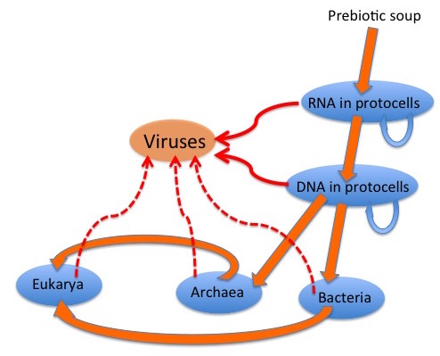
The graphic depicts a possible scenario for the evolution of viruses: “wild” genetic elements could have escaped, or even been the agents for transfer of genetic information between, both RNA-containing and DNA-containing “protocells”, to provide the precursors of retroelements and of RNA and DNA viruses.  Later escapes from Bacteria, Archaea and their progeny Eukarya would complete the virus zoo.

virus descent

It is generally accepted that many viruses have their origins as “escapees” from cells; rogue bits of nucleic acid that have taken the autonomy already characteristic of certain cellular genome components to a new level.  Simple RNA viruses are a good example of these: their genetic structure is far too simple for them to be degenerate cells; indeed, many resemble renegade messenger RNAs in their simplicity.

RdRp cassettes and virus evolution

RNA virus supergroups and RdRp and CP cassettes

What they have in common is a strategy which involves use of a virus-encoded RNA-dependent RNA polymerase (RdRp) or replicase to replicate RNA genomes – a process which does not occur in cells, although most eukaryotes so far investigated do have RdRp-like enzymes involved in regulation of gene expression and resistance to viruses.  The surmise is that in some instances, an RdRp-encoding element could have became autonomous – or independent of DNA – by encoding its own replicase, and then acquired structural protein-encoding sequences by recombination, to become wholly autonomous and potentially infectious.

A useful example is the viruses sometimes referred to as the “Picornavirus-like” and “Sindbis virus-like” supergroups of ssRNA+ viruses, respectively.  These two sets of viruses can be neatly divided into two groups according to their RdRp affinities, which determine how they replicate.  However, they can also be divided according to their capsid protein affinities, which is where it is obvious that the phenomenon the late Rob Goldbach termed “cassette evolution” has occurred: some viruses that are relatively closely related in terms of RdRp and other non-structural protein sequences have completely different capsid proteins and particle morphologies, due to acquisition by the same RdRp module of different structural protein modules.

Given the very significant diversity in these sorts of viruses, it is quite possible that this has happened a number of times in the evolution of cellular organisms on this planet – and that some single-stranded RNA viruses like bacterial RNA viruses or bacteriophages and some plant viruses (like Tobacco mosaic virus, TMV) may be very ancient indeed.

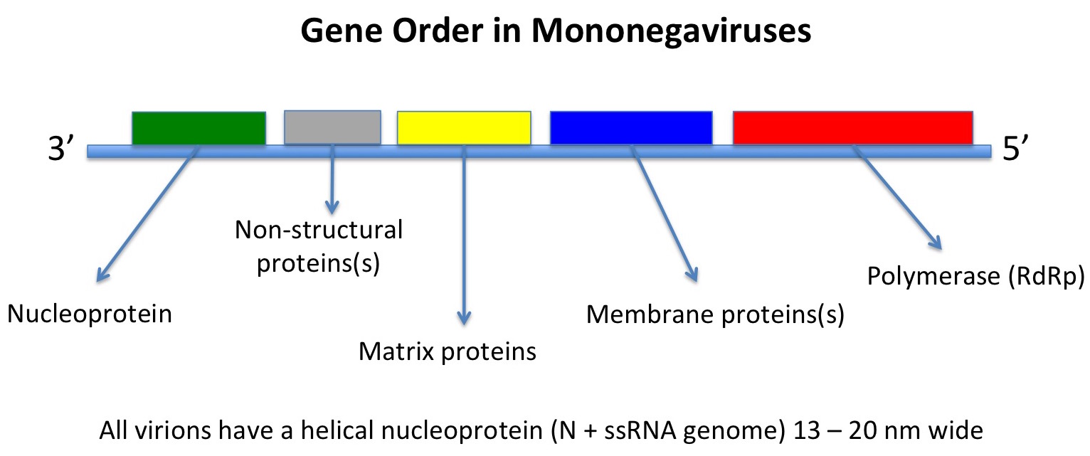
However, other ssRNA viruses – such as the negative sense mononegaviruses, Order Mononegaviraleswhich includes the families Bornaviridae, Rhabdoviridae, Filoviridae and Paramyxoviridae, represented by Borna disease virus, rabies virus, Zaire Ebola virus, and measles and mumps viruses respectively – may be evolutionarily much younger.  In this latter case, the viruses all have the same basic genome with genes in the same order and helical nucleocapsids within differently-shaped enveloped particles.

Their host ranges also indicate that they originated in insects: the ones with more than one phylum of host either infect vertebrates and insects or plants and insects, while some infect insects only, or only vertebrates – indicating an evolutionary origin in insects, and a subsequent evolutionary divergence in them and in their feeding targets.

Slide1

HIV: a retrovirus

The Retroid Cycle

The ssRNA retroviruses – like HIV – are another good example of possible cell-derived viruses, as many of these have a very similar genetic structure to elements which appear to be integral parts of cell genomes – termed retrotransposons –  and share the peculiar property of replicating their genomes via a pathway which goes from single-stranded RNA through double-stranded DNA (reverse transcription) and back again, and yet have become infectious.  They can go full circle, incidentally, by permanently becoming part of the cell genome by insertion into germ-line cells – so that they are then inherited as “endogenous retroviruses“, which can be used as evolutionary markers for species divergence.

The Retroid Cycle

Indeed, there is a whole extended family of reverse-transcribing mobile genetic elements in organisms ranging from bacteria all the way through to plants, insects and vertebrates, indicating a very ancient evolutionary origin indeed – and which includes two completely different groups of double-standed DNA viruses, the vertebrate-infecting hepadnaviruses or hepatitis B virus-like group, and the plant-infecting badna- and caulimoviruses.

Metaviruses and pseudoviruses

These are two families of long terminal repeat-containing (LTR) retrotransposons, with different genetic organisations. 

Members of family Pseudoviridae, also known as Ty1/copia elements,  have polygenic genomes of 5-9 kb ssRNA which encode a retrovirus-like Gag-type protein, and a polyprotein with protease (PR), integrase (IN) and reverse transcriptase / RNAse H  (RT) domains, in that order.  While some members also encode an env-like ORF, the 30-40 nm particles that are an essential replication intermediate have no envelope or Env protein.  They are not infectious.  Host species include yeasts, insects, plants and algae.

Metaviruses – family Metaviridae – are also known as Ty3-gypsy elements, and have ssRNA genomes of 4-10 kb in length.  They replicate via particles 45-100 nm in diameter composed of Gag-type protein, and some species have envelopes and associated Env proteins.  Gene order in the genomes is Gag-PR-RT-IN-(Env), as for retroviruses.  One virus – Drosophila melanogaster Gypsy virus – is infectious; however, as for pseudoviruses, most are not.  The genomes have been found in all lineages of eukaryotes so far studied in sufficient detail.

Both pseudovirus and metavirus genomes are clearly related to classic retroviruses; moreover, RT sequences point to metavirus RTs being most closely related to plant DNA pararetrovirus lineage of caulimoviruses.  This gives rise to the speculation that pseudoviruses and metaviruses have a common and ancient ancestor – and that two different metavirus lineages gave rise to retroviruses and caulimoviruses respectively.

All of these cellular elements and viruses have in common a “reverse transcriptase” or RNA-dependent DNA polymerase, which may in fact be an evolutionary link back to the postulated “RNA world” at the dawn of evolutionary history, when the only extant genomes were composed of RNA, and probably double-stranded RNA.  Thus, a part of what could be a very primitive machinery indeed has survived into very different nucleic acid lineages, some viral and many wholly cellular in nature, from bacteria through to higher eukaryotes.

The possibility that certain non-retro RNA viruses can actually insert bits of themselves by obscure mechanisms into host cell genomes – and afford them protection against future infection – complicates the issue rather, by reversing the canonical flow of genetic material.  This may have been happening over aeons of evolutionary time, and to have involved hosts and viruses as diverse as plants (integrated poty– and geminivirus sequences), honeybees (integrated Israeli bee paralysis virus) – and the recent discovery of “…integrated filovirus-like elements in the genomes of bats, rodents, shrews, tenrecs and marsupials…” which, in the case of mammals, transcribed fragments “…homologous to a fragment of the filovirus genome whose expression is known to interfere with the assembly of Ebolavirus”.

Rolling circle replication

There are also obvious similarities in mode of replication between a family of elements which include bacterial plasmids, bacterial single-strand DNA viruses, and viruses of eukaryotes which include geminiviruses and nanoviruses of plants, parvoviruses of insects and vertebrates, and circoviruses and anelloviruses of vertebrates.

Geminivirus particle

These agents all share a “rolling circle” DNA replication mechanism, with replication-associated proteins and DNA sequence motifs that appear similar enough to be evolutionarily related – and again demonstrate a continuum from the cell-associated and cell-dependent plasmids through to the completely autonomous agents such as relatively simple but ancient bacterial and eukaryote viruses.

geminivirus rolling circle replication

Big DNA viruses

Mimivirus particle, showing basic structure

However, there are a significant number of viruses with large DNA genomes for which an origin as cell-derived subcomponents is not as obvious.  In fact, one of the largest viruses yet discovered – mimivirus, with a genome size of greater than 1 million base pairs of DNA – have genomes which are larger and more complex than those of obligately parasitic bacteria such as Mycoplasma genitalium (around 0.5 million), despite their sharing the life habits of tiny viruses like canine parvovirus (0.005 million, or 5000 bases).

Mimivirus has been joined, since its discovery in 2003, by Megavirus (2011; 1.2 Mbp) and now Pandoravirus (2013; 1.9 -2.5 Mbp). 

The nucleocytoplasmic large DNA viruses or NCLDVs – including pox-, irido-, asfar-, phyco-, mimi-, mega- and pandoraviruses, among others – have been grouped as the proposed Order Megavirales, and it is proposed that they evolved, and started to diverge, before the evolutionary separation of eukaryotes into their present groupings.

It is a striking fact that the largest viral DNA genomes so far characterised seem to infect primitive eukaryotes such as amoebae and simple marine algae – and they and other large DNA viruses like pox- and herpesviruses seem to be related to cellular DNA sequences only at a level close to the base of the “tree of life”.

Variola virus, the agent of smallpox. Image courtesy Russell Kightley Media.

This indicates a very ancient origin or set of origins for these viruses, which may conceivably have been as obligately parasitic cellular lifeforms which then made the final adaptation to the “virus lifestyle”.

However, their actual origin could be in an even more complex interaction with early cellular lifeforms, given that viruses may well be responsible for very significant episodes of evolutionary change in cellular life, all the way from the origin of eukaryotes through to the much more recent evolution of placental mammals.  In fact, there is informed speculation as to the possibility of viruses having significantly influenced the evolution of eukaryotes as a cognate group of organisms, including the possibility that a large DNA virus may have been the first cellular nucleus.

In summary, viruses are as much a concept as a unitary entity: all viruses have in common, given their polyphyletic origins, is a base-level strategy for replicating their genomes.  Otherwise, their origins are possibly as varied as their genomes, and may remain forever obscure.

I am indebted to Russell Kightley for use of his excellent virus images.

Updated 12th August 2015