Posts Tagged ‘vaccine’

Nano Patents and Innovations: Powerful New Approach To Attack Flu Virus

28 May, 2012

See on Scoop.itVirology and Bioinformatics from Virology.ca

An international research team has manufactured a new protein that can combat deadly flu epidemics.

The paper, featured on the cover of the current issue of Nature Biotechnology, demonstrates ways to use manufactured genes as antivirals, which disable key functions of the flu virus, said Tim Whitehead, assistant professor of chemical engineering and materials science at Michigan State University.

See on nanopatentsandinnovations.blogspot.fr

ProMED-mail | MEASLES UPDATE 2012 (22)

28 May, 2012

See on Scoop.itVirology and Bioinformatics from Virology.ca

ProMED Mail is possibly THE premier infectious disease updating service in the world today, having sprung to fame during the Kikwit Ebola outbreak in 1995.  This is one of a series of posts on measles, which is documenting a very disturbing trend: the incidence of the disease is increasing in places where it should have been eradicated, because well-educated and sophisticated communities are not vaccinating their children – or themselves.


One very telling quote from the post:

“Measles is highly infectious so we must all do
everything possible to prevent the spread of it, particularly with an outbreak on our doorstep. … MMR vaccination is the only way to prevent
measles. If parents haven’t arranged for their children to be vaccinated – it’s not too late to have the jab. Parents don’t realise that measles is not just a case of a few spots – it can be a very serious illness. Symptoms include fever, cough, soreness of the eyes and a rash which spreads rapidly over the body. Serious complications affect one in 15 children. These include chest infections, fits, encephalitis (swelling of the brain), and brain damage. In very serious cases, measles can kill.”

See on www.promedmail.org

Pandemic 2009 H1N1 vaccination produces antibodies against multiple flu strains

27 May, 2012

See on Scoop.itVirology News

“The pandemic 2009 H1N1 vaccine can generate antibodies in vaccinated individuals not only against the H1N1 virus, but also against other influenza virus strains including H5N1 and H3N2.”


And a possible reason for this could be that the H1N1pdm virus’ haemagglutinin is a natural “ancestral” sequence – the kind that HIV vaccine researchers are looking for for gp120/160, which have been shown to elicit a wider spectrum of cross-reacting antibodies than “evolved” proteins, or ones that have been selected for antigenic escape in humans for a good few viral generations.


Flu vaccine graphic by Russell Kightley Media

See on www.eurekalert.org

Trends in Intussusception Hospitalizations Among US Infants Before and After Implementation of the Rotavirus Vaccination Program, 2000–2009

27 May, 2012

See on Scoop.itVirology News

“A small increase in intussusception rates was seen among infants aged 8–11 weeks, to whom most first doses of rotavirus vaccine were given, but no sustained population-level change in overall intussusception hospitalizations rates in US infants was observed after implementation of the US rotavirus vaccination program. Although an association between intussusception and rotavirus vaccination cannot be established by this ecologic analysis alone, even if the low risk with the first dose exists, it is outweighed by the well-documented benefits of vaccination of US infants”

This is a big deal- a very important, big deal: human rotavirus kills more than 500 000 people a year (mainly very little), and rotavirus vaccines have been bedevilled with the suspicion that they cause telescoping of the intestine, or intussusception.  Which can be fatal, and is not something you want happening to your healthy baby.

However, and however: I have taught my students for years to be aware of relative risks when talking about vaccines, and there is absolutely no doubt that even the Wyeth vaccine could have been considered “safe” in a developing country environment, where the threat of death due to diarrhoea and dehyderation caused by rotavirus, would have been far greater than any threat from the vaccine.

I thank Rusdsell Kightley Media for the rotavirus graphic

See on jid.oxfordjournals.org

Flu shot offers surprising benefits for pregnant women; Vaccine may fight stillbirth, preterm birth, and very low birth weight

25 May, 2012

See on Scoop.itVirology News

A new study announced Tuesday finds the H1N1 flu vaccine not only can protect you from getting sick but can actually benefit your baby.

Researchers from the University of Ottawa in Canada examined data from more than 55,000 child births in Ontario during an outbreak of H1N1, comparing mothers who were vaccinated to those who weren’t.

While prior research has found that pregnant women can safely get the flu shot at any stage of their pregnancies — something many doctors vehemently support — the new findings associate H1N1 vaccinations with a significantly reduced risk of stillbirth, preterm birth, and very low birth weight.

“These are all significant results, but especially interesting is the finding that the vaccinated mothers were one-third less likely to have a stillborn child,” said study researcher Deshayne Fell, a graduate student at McGill University who works with the birth record database. “This is one of the only studies large enough to evaluate the association between maternal flu vaccination and stillbirth — a very rare event.”


So much for the disinformation about dangers to pregnant women: in fact, Spanish Flu and the recent H1N1 pandemic were both especially dangerous for unprotected pregnant women.

See on www.nydailynews.com

Muslim leaders enlisted to help stamp out polio

25 May, 2012

See on Scoop.itVirology News

GENEVA (Reuters) – The last three countries where polio is still paralyzing children — Afghanistan, Pakistan and Nigeria — said on Thursday that they have enlisted Muslim women and religious leaders to allay fears of vaccination and wipe out the disease.
Polio cases are at an all-time low worldwide, following its eradication in India last year, raising hopes but also fears about a threat of resurgence especially in sub-Saharan Africa unless remaining reservoirs of polio virus are stamped out.

Conflict and insecurity is preventing health workers from reaching hundreds of thousands of children in Afghanistan and Pakistan with doses of polio vaccine, health ministers said.

See on www.reuters.com

Engineered H5N1: the wheels grind on, and on, and on….

19 April, 2012

The Scientist has a nice collection of articles on this topic, which I have commented on all over the place, so I though I might consolidate some of it in one place.

In response to the article entitled “Deliberating Over Danger“, I wrote the following:

The point I and others have made before is that H5N1 and other influenza viruses are not waiting for us to let engineered versions loose, before they cause pandemics: all of the mutations noted by the Fouchier and Kawaoka groups are almost certainly present in the several environments where H5N1 viruses are now endemic – and all it takes for all of them to be present together is a little more mixing.

Don’t discount other flu subtypes, either: while everyone is obsessing about H5N1, H3N2 is busy popping out of pigs in the USA; H9N2 in birds in Bangladesh; H5N2 in ostriches in South Africa – and all it would take is one or a couple of fortuitous reassortments, and a whole new flu virus could be unleashed.

While the “deadly” H5N1s are being worked on in lockdown facilities.

If we don’t know what the virus does, we won’t know what it can do. If we don’t know what to look for, we may be taken unawares, when the next 1918-type pandemic strikes.

I want to have universal flu vaccines by then – so we won’t HAVE to worry about a new flu

.

There are also three newer articles covering the controversy: these are

  • H5N1 Researcher to Defy Dutch Gov’t?
  • (with my comment – “Export permit to publish something?  Really?  A complete misapplication of laws to material that should not be subject to them.”)
  • White House Weighs in on H5N1
  • Flu Review Criticized
  • (with my comment – “So after a full and frank hearing did not go his way, after changes had been made to the paper in question (Fouchier’s), Osterholm complains.  Such is life….”

There is the slightly older article – “Bird Flu Papers to Publish” – describing the reversal of the NSABB’s decision to ask for redaction of the two papers describing mammal-to-mammal aerosol-transmissible H5N1.

An interesting article also describes Yoshihiro Kawaoka’s results:

“First, he introduced two mutations—N224K and Q226L—into the haemagglutinin (HA) protein of H5N1 that made the virus capable of sticking to receptors on human tracheal cells. Then he created a chimeric virus by combining the mutated HA protein with genes from the H1N1 virus, which sparked a pandemic in 2009. Kawaoka identified another HA mutation, called N158D, that allowed the virus to spread between ferrets that were not in direct physical contact. A fourth mutation, T318I, also showed up in the H5N1 strain, but its role in making the virus more transmissible among mammals is less clear.”

So there you are: an actual recipe for aerosol-transmissible H5N1.  It was always going to come out somehow, and now these two papers will probably the most cited flu papers ever.  Nothing like a little hype!  Meanwhile, H5 and its brothers and sisters are out there mutating away, with no help needed from anyone.  Roll on universal flu vaccines!!

The origin of HIV: still so much garbage out there

20 February, 2012

While curating Virology News today, I came across another reprocessing of new that I had come across earlier concerning apparent natural protection of some African female sex workers against HIV infection: this was the intriguingly-entitled “African women’s genitals provide clue to HIV prevention“, in what appears to be an online Nigerian newspaper.

This recapitulates, very accurately, the information I reported in Virology News, which was the subject of a news release following the publication in the September 2011 edition of PLoS One of a study entitled “High Level of Soluble HLA-G in the Female Genital Tract of Beninese Commercial Sex Workers Is Associated with HIV-1 Infection”.  The gist of this is that:

“HIV-resistant sex workers in Africa have a weak inflammatory response in their vaginas – a surprise for the researchers, who were expecting the contrary considering the women’s high exposure to the virus.”

This may lend further credence to the observation that progression to AIDS in HIV-infected people is associated with a state of chronic immune activation – and that SIV-infected vervet monkeys do not exhibit such chromic immune activation, and do not progress like humans do.

What is interesting about the Nigerian article, however, is not what it reports – it is the online comments that follow it.  Here is a selection:

“Was HIV realy discovered in Africa ? Forget the western media propaganda . I have believed , for years , that HIV is a laboratory virus designed for genocide in the thick of apartheid inhuman policies in South Africa .”

“Neither did HIV originate  nor was it perculiar to Africa. It was the creation of the Western countries to stsyematically reduce African population. that the subjects of this study were exposed to HIV virus attests to this fact.”

And my personal favourite:

“So you have already swallowed up the white propaganda that the AIDS virus was first discovered in 1981 in a remote area of central Africa in the green monkey!  A fairy tale, which never explains why prior to its first clinical detection among western homosexual men in the late seventies, no case was found in Africans, and no animal or human population died off in Africa, yet the homosexual population of the west was seriously threatened until their protected sex campaign took off.

You must be unaware that about 35 years ago the Soviet KGB told the world the truth about AIDS….

Jakob Segal, a former biology professor at Humboldt University in then-East Germany, proposed that HIV was engineered at a U.S. military laboratory at Fort Detrick, by splicing together two other viruses, Visna and HTLV-1. According to his theory, the new virus, created between 1977 and 1978, was tested on prison inmates who had volunteered for the experiment in exchange for early release. He further suggested that it was through these prisoners, most of who were homosexuals, that the virus was spread to the population at large.”

What is depressing is that there is just one comment saying “…where HIV started is of little significance now. the issue is that our brothers Africans are the ones affected so we must work hard to find the cure and save our brothers.”

What is obvious is that, even in an environment such as one of the most developed nations in Africa, where intelligent science reporting is happening, the public seems to be alarmingly misinformed about the origin of HIV and predisposed to believe racist conspiracy theories that were debunked years ago.

FACT:
HIV did not come from “green monkeys” and was not discovered in 1981: the virus was described in 1983 and 1984, and HIV entered the  human population in central Africa multiple times, from chimpanzees and possibly also from gorillas, almost certainly via bushmeat – and this happened in the 1930s or even earlier.

FACT:
HIV could not possibly have resulted  from the splicing together of Visna virus and HTLV-1, as no HIV sequence bears any strong resemblance to either virus – and especially not to both of them in different parts of their genomes, as they would be expected to if they were artificial recombinants.  Moreover, the first HIV that has been reliably dated comes from a sample taken in the Congo in 1959.

All of these facts can be easily discovered by a trawl of either the scientific literature, or a first-level digest of that literature by reputable journalists.  All else is fiction…and malicious fiction at that, whether or not such supposed luminaries as Thabo Mbeki believe it.

12th May 2015

ANOTHER note added in response to Timothy Julian, below, who seems not to understand anything about retrovirus and especially lentivirus evolution.  Here is an unrooted radial relationship diagram (aka “phylogenetic” diagram) depicting whole genome sequence relationships between HIV-1, HIV-2, 2 SIVs, Maedi-Visna ad bovine leukaemia viruses, feline and bovine immunodeficiency and human and simian T-cell lymphotropic viruses.  Done by me today from Genbank sequences, using CLC Genomics Workbench ver 7.

Radial tree for retrovirus complete genome sequences

Radial tree for retrovirus complete genome sequences

What it shows is that:

  • there is a distinctive clustering of HIVs and of SIVs, with MVV as a apparently closer relative than the FIVs, in a cluster of lentiviruses that includes BIV – and I note HIV-1 is more closely related to an SIV than it is to HIV-2, and there are three branches to the H/SIV tree ALL of which are internal to MVV and the FIVs and BIV.
  • the H/STLVs cluster together as relatives, with HTLV-1/2/3 being most closelt related to STLV-1/2/3 – which, seeing as the HTLVs are supposed to have derived from the STLVs, is hardly surprising.
  • BLV is only distantly related to the TLV cluster, as is expected given that it is a leukaemia virus but one of a very different species

If HIV-1 derives from artificial constructs derived from FIVs, which are less closely related to  them than is MVV, then is the same true for the whole primate cluster?  Really?  When it is pretty obvious that they are (a) evolutionarily related most closely to one another, (b) evolutionarily diverged to quite a considerable extent?  So were they all made individually??  Then cleverly given to different bush-dwelling primates in Africa?  How desperately unlikely is that??  You appear not to have heard of teh principle of parsimony, which is that the simplest explanation that covers all of the facts is probably correct – which in this case, is that both HIVs and all of the SIVs have a common evolutionary origin, thousands of years ago – and that all lentiviruses also have a common origin, millions of years ago.

Seriously, Timothy: give it a rest.  You know less than Jon Snow.

And while they were arguing about killer H5N1…

8 February, 2012

…Elsevier’s Virology was calmly publishing another paper on a “mutant” H5N1….

The abstract:

Acquisition of α2-6 sialoside receptor specificity by α2-3 specific highly-pathogenic avian influenza viruses (H5N1) is thought to be a prerequisite for efficient transmission in humans. By in vitro selection for binding α2-6 sialosides, we identified four variant viruses with amino acid substitutions in the hemagglutinin (S227N, D187G, E190G, and Q196R) that revealed modestly increased α2-6 and minimally decreased α2-3 binding by glycan array analysis. However, a mutant virus combining Q196R with mutations from previous pandemic viruses (Q226L and G228S) revealed predominantly α2-6 binding. Unlike the wild type H5N1, this mutant virus was transmitted by direct contact in the ferret model although not by airborne respiratory droplets. However, a reassortant virus with the mutant hemagglutinin, a human N2 neuraminidase and internal genes from an H5N1 virus was partially transmitted via respiratory droplets. The complex changes required for airborne transmissibility in ferrets suggest that extensive evolution is needed for H5N1 transmissibility in humans. [my emphasis – Ed]

I have covered the use of glycan arrays to characterise influenza viruses’ binding specificity previously; I thought then, and do now, that it is a very cool technology – and one that has shown in this case that H5N1 variants can be selected from an originally “wild” population, that preferentially bind the human-type receptor.

And they did it like this:

To examine the functional evolution of H5 HA receptor specificity in the laboratory, we implemented an in vitro receptor-binding virus enrichment approach that recapitulates in vivo selection. Synthetic 6′-sialyl (N-acetyl-lactosamine) (6′ SLN) was used as the affinity ligand mimicking the human receptor to capture spontaneous viral receptor variants on the surface of magnetic beads. Starting with a pool of 108 EID50 of A/Vietnam/1203/2004 (VN04 virus), we performed four consecutive rounds of in vitro binding and elution followed by isolation of 150 individual virus clones by plaque purification and characterization by sequence analysis.

No “genetic engineering” here – or furore over “killer viruses escaping the lab!”  Possibly because (a) “mutant virus was transmitted by direct contact in the ferret model although not by airborne respiratory droplets”, and (b) “a reassortant virus with the mutant hemagglutinin, a human N2 neuraminidase and internal genes from an H5N1 virus was partially transmitted via respiratory droplets” [my emphasis].

Meaning they didn’t actually make anything that could immediately elicit such scare-mongering as the more notorious studies I and many others have reported on previously.

However, the grim NSABB folk were quick to decry the publication, saying “”I think it is fair to say that we would have liked to have seen it before it was published,” [Paul Keim, chairman of the National Science Advisory Board for Biosecurity], and the “…altered bird flu virus could mutate in dangerous ways if unleashed in nature”.

I am more worried, to be perfectly honest, over the dangerous ways the the wild type virus could mutate IN nature, given that mutants can be selected so apparently easily!

A Short History of the Discovery of Viruses – Part 2

7 February, 2012

The following text has now appeared in modified form in an ebook, for sale for US$4.99 on the iBooks Store

The Ultracentrifuge, Eggs and Flu

The ultracentrifuge

A technical development that was to greatly advance the study of viruses was begun in 1923, but only reached fruition by the 1930s: this was the ultracentrifuge, invented and developed first by Theodor (“The”) Svedberg in Sweden as a purely analytical tool, and later by JW Beams and EG Pickels in the USA as an analytical and preparative tool.  The ultracentrifuge revolutionised first, the physical analysis of proteins in solution, and second, the purification of proteins, viruses and cell components, by allowing centrifugation at speeds high enough to allow pelleting of subcellular fractions.

Analytical centrifugation and calculation of molecular weights of particles gave some of the first firm evidence that certain proteins, and virus particles, were large, regular objects.  Indeed, it came to be taken as a given that one of the fundamental properties of a virus particle was its sedimentation coefficient, measured in svedbergs (a unit of 10-13 seconds, shown as S20,W).  This is also how ribosomes of pro- and eukaryotes came to be named: these are known as 70S (prokaryote) and 80S ribosomes, respectively, based on their different sedimentation rates.

The Official Discovery of Influenza Virus

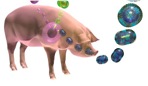
In 1931, Robert Shope in the USA managed to recreate swine influenza by intranasal administration of filtered secretions from infected pigs.  Moreover, he showed that the classic severe disease required co-inoculation with a bacterium – Haemophilus influenza suis – originally thought to be the only agent.  He also pointed out the similarities between the swine disease and the Spanish Flu, where most patients died of secondary infections.  However, he also suggested that the virus survived seasonally in a cycle involving the pig, lungworms, and the earthworm, which is now known to be completely wrong.

This notwithstanding, he found that people who had survived infection during the 1918 pandemic had antibodies protecting them against the swine flu virus, while people born after 1920 did not, which showed that the 1918 human and swine flu viruses were very similar if not identical. This was a very relevant discovery for what happened much later, in the 2009 influenza pandemic, when the same virus apparently came back into the human population from pigs after circulating in them continuously since 1918.

Shope went on in 1932 to discover, with Peyton Rous, what was first called the Shope papillomavirus and later Cottontail rabbit papillomavirus: this causes benign cancers in the form of long hornlike growths on the head and face of the animal. This may explain the sightings in the US Southwest of the near-mythical “jackalope”.

Influenza viruses in pigs

Influenza viruses in pigs

Patrick Laidlaw and William Dunkin, working in the UK at the National Institute for Medical Research (NIMR), had by 1929 successfully characterised the agent of canine distemper – a relative of measles, mumps and distemper morbilliviruses – as a virus, proved it infected dogs and ferrets, and in 1931 got a vaccine into production that protected dogs.  This was made from chemically inactivated filtered tissue extract from infected animals.  Their work built on and completely eclipsed earlier findings, such as those of Henri Carré in France in 1905, who first claimed to have shown it was a filterable agent, and Vittorio Puntoni, who first made a vaccine in Italy from virus-infected brain tissue inactivated with formalin in 1923.

Influenza and Ferrets: the Early Days

Continuing from Laidlaw and Dunkin’s work in the same institute, Christopher Andrewes, Laidlaw and W Smith reported in 1933 that they had isolated a virus from humans infected with influenza from an epidemic then raging.  They had done this by infecting ferrets with filtered extracts from infected humans – after the fortuitous observation that ferrets could apparently catch influenza from infected investigators!  The “ferret model” was very valuable – see here for modern use of ferrets – as strains of influenza virus could be clinically distinguished from one another.

Eggs and Flu and Yellow Fever

Influenza virus and eggs: large-scale culture

Frank Macfarlane Burnet from Australia visited the NIMR in the early 1930s, and learned a number of techniques he used to great effect later on.  Principal among these was the technique of embryonated egg culture of viruses – which he took back to Melbourne, and applied to the infectious laryngotracheitis virus of chickens in 1936.  This is a herpesvirus, first cultivated by JR Beach in the USA in 1932: Burnet used it to demonstrate that it was possible to do “pock assays” on chorioallantoic membranes that were very similar to the plaque assays done for bacteriophages, with which he was also very familiar.  Also in 1936, Burnet started a series of experiments on culturing human influenza virus in eggs: he quickly showed that it was possible to do pock assays for influenza virus, and that

“It can probably be claimed that, excluding the bacteriophages, egg passage influenza virus can be titrated with greater accuracy than any other virus.”

Max Theiler and colleagues in the USA took advantage of the new method of egg culture to adapt the French strain of yellow fever virus (YFV) he had grown in mouse brains to being grown in chick embryos, and showed that he could attenuate the already weakened strain even further – but it remained “neurovirulent”, as it caused encephalitis or brain inflammation in monkeys.  He then adapted the first YFV characterised – the Asibi strain, from Ghana in 1927 – to being grown in minced chicken embryos lacking a spinal cord and brain, and showed in 1937 that after more than 89 passages, the virus was no longer “neurotrophic”, and did not cause encephalitis.   The new 17D strain of YFV was successfully tested in clinical trials in Brazil in 1938 under the auspices of the Rockefeller Foundation, which has supported YFV work since the 1920s.  The strain remains in use today, and is still made in eggs.

Virus purification and the physicochemical era

Given that the nature of viruses had prompted people to think of them as “chemical matter”, researchers had attempted from early days to isolate, purify and characterise the infectious agents.  An early achievement was the purification of a poxvirus in 1922 by FO MacCallum and EH Oppenheimer. 

Much early work was done with bacteriophages and plant viruses, as these were far easier to purify or extract at the concentrations required for analysis, than animal or especially human viruses. 

CG Vinson and AM Petre, working with the infectious agent causing mosaic disease in tobacco – tobacco mosaic virus, or TMV – showed in 1931 that they could precipitate the virus from suspension as if it were an enzyme, and that infectivity of the precipitated preparation was preserved.  Indeed, in their words:

“…it is probable that the virus which we have investigated reacted as a chemical substance”.

Viruses in Crystal

An important set of discoveries started in 1935, when Wendell Stanley in the USA published the first proof that TMV could be crystallised, at the time the most stringent way of purifying molecules.  He also reported that the “protein crystals” were contaminated with small amounts of phosphorus.  An important finding too, using physical techniques including ultracentrifugation and later, electron microscopy, was that the TMV “protein” had a very high molecular weight, and was in fact composed of large, regular particles.  This was a very significant discovery, as it indicated that some viruses at least really were very simple infectious agents indeed.

TMV particle: 95% protein, 5% RNA

However, his conclusion that TMV was composed only of protein was soon challenged, when Norman Pirie and Frederick Bawden working in the UK showed in 1937 that ribonucleic acid (RNA) – which consists of ribose sugar molecules linked by phosphate groups – could be isolated consistently from crystallised TMV as well as from a number of other plant viruses, which accounted for the phosphorus “contamination”.  This resulted in the realisation that TMV and other plant virus particles – now known to be virions – were in fact nucleoproteins, or protein associated with nucleic acid.

Stanley received a share of the Nobel Prize in Chemistry in 1946 for his work on TMV: it is instructive to read his acceptance speech from the time to realise what the state of the science that was becoming virology was at the time.  He wrote:

“Since the original discovery of this infectious, disease-producing agent, known as tobacco mosaic virus, well over three hundred different viruses capable of causing disease in man, animals and plants have been discovered. Among the virus-induced diseases of man are smallpox, yellow fever, dengue fever, poliomyelitis, certain types of encephalitis, measles, mumps, influenza, virus pneumonia and the common cold. Virus diseases of animals include hog cholera, cattle plague, foot-and-mouth disease of cattle, swamp fever of horses, equine encephalitis, rabies, fowl pox, Newcastle disease of chickens, fowl paralysis, and certain benign as well as malignant tumors of rabbits and mice. Plant virus diseases include tobacco mosaic, peach yellows, aster yellows, potato yellow dwarf, alfalfa mosaic, curly top of sugar beets, tomato spotted wilt, tomato bushy stunt, corn mosaic, cucumber mosaic, and sugar cane yellow stripe. Bacteriophages, which are agents capable of causing the lysis of bacteria, are now regarded as viruses”.

Two of the most interesting things about the article, however, are the electron micrographs of virus particles – Stanley had one of the first electron micrsoscopes available at the time –  and the table of sizes of viruses, proteins and cells that had been determined by then by techniques such as ultracentrifugation and filtration: TMV was known to be rodlike, 15 x 280 nm; vaccinia was 210 x 260 nm; poliomyelitis was 25 nm; phages like T2 were known to have a head-and-tail structure.

Seeing is Believing: the Electron Microscope

First Electron Microscope with Resolving Power Higher than that of a Light Microscope. Ernst Ruska, Berlin 1933 Wikipedia CC BY-SA 3.0, https://www.flickr.com/photos/93452909@N00/176059674

First Electron Microscope with Resolving Power Higher than that of a Light Microscope. Ernst Ruska, Berlin 1933
Wikipedia CC BY-SA 3.0, https://www.flickr.com/photos/93452909@N00/176059674

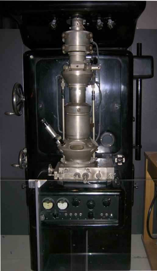
The development of the electron microscope, in Germany in the 1930s, represented a revolution in the investigation of virus structures: while virions of viruses like variola and vaccinia could just about be seen by light microscopy – and had been, as early as 1887 by John Buist and others – most viruses were far too small to be visualised in this way. 

While Ernst Ruska received a Nobel Prize in 1986 for developing the electron microscope, it was his brother Helmut who first imaged virus particles – using beams of electrons deflected off virus particles coated in heavy metal atoms.  From 1938 through the early 1940s, using his “supermicroscope”, he imaged virions of poxviruses, TMV, varicella-zoster herpesvirus, and bacteriophages, and showed that they were all particulate – that is, they consisted of regular and sometimes complex particles, and were often very different from one another.  He even proposed in 1943 a system of viral classification on the basis of their perceived structure.

While electron microscopy was also used medically to some extent thereafter – for example, in differentiating smallpox from chickenpox by imaging particles of variola virus and varicella-zoster virus, respectively, derived from patients’ vesicles – its use was limited by the expense and cumbersome nature of sample preparation. For example, the micrographs in Stanley’s 1946 paper were all done with samples “…prepared with gold by the shadow-casting technique”.

The use of the cumbersome technique of metal shadow-casting, and the highly inconvenient nature of electron microscopy as a routine tool all changed from 1959 onwards, when Sydney Brenner and Robert Horne published “A negative staining method for high resolution electron microscopy of viruses”.  This method involves the use of viruses in liquid samples deposited on carbon-coated metal grids, and then stained with heavy-metal salts such as phosphotungstic acid (PTA) or uranyl acetate.

This simple technique revolutionised the field of electron microscopy, and within just a few years much information was acquired about the architecture of virus particles. Not only were the overall shapes of particles revealed, but also the details of the symmetrical arrangement of their components. Some beautiful examples can be seen here, at the Cold Spring Harbor site.

Depiction of the effects of using a heavy metal salt solution to negatively stain particles on a carbon film. The stain (dark) pools around the particles (light).  Human rotavirus particles, stained from below (left) and by immersion (right).
Images copyright LM Stannard

Depiction of the effects of using a heavy metal salt solution to negatively stain particles on a carbon film. The stain (dark) pools around the particles (light). Human rotavirus particles, stained from below (left) and by immersion (right).
Images copyright LM Stannard

Click here for Part 1: Filters and Discovery

here for Part 3: Phages, Cell Culture and Polio

and here for Part 4: RNA Genomes and Modern Virology

Copyright Edward P Rybicki and Russell Kightley, February 2015, except where otherwise noted.