Archive for the ‘Vaccines: General’ Category

PBVAB 5 – Part 3

21 August, 2013

PBVAB 5 Verona, June 2013 – Part 3

Technically, Sue Huddy’s piece should have been Part 3; however, it reports things that happened after what I am reporting on, so I’ll keep that label!

This post will report on Sessions 3 & 4, namely, Technology Advances and Perspectives.

I opened Session 3 with a talk on ‘Virus-derived ssDNA vectors for the expression of foreign proteins in plants’, focusing mainly on geminiviruses (naturally).  I wrote this a couple of years ago as a chapter for a book which seemed to not be forthcoming; however, I was assured during my talk by Yuri Gleba – the co-Editor with Kenneth Palmer of a “Current Topics in Microbiology and Immunology” issue on “Plant Viral Vectors” – that this offering is now in fact available, so here’s a link for anyone who wants to buy it.

Current Topics in Microbiology and Immunology 2011,

Virus-Derived ssDNA Vectors for the Expression of Foreign Proteins in Plants

Edward P. RybickiDarrin P. Martin

Plant viruses with ssRNA genomes provide a unique opportunity for generating expression vehicles for biopharming in plants, as constructs containing only the replication origin, with the replication-associated protein (Rep) gene provided in cis or in trans, can be replicationally amplified in vivo by several orders of magnitude, with significant accompanying increases in transcription and expression of gene(s) of interest. Appropriate replicating vectors or replicons may be derived from several different generic geminiviruses (family Geminiviridae) or nanoviruses (family Nanoviridae), for potential expression of a wide range of single or even multiple products in a wide range of plant families. The use of vacuum or other infiltration of whole plants by Agrobacterium tumefaciens suspensions has allowed the development of a set of expression vectors that rival the deconstructed RNA virus vectors in their yield and application, with some potential advantages over the latter that still need to be explored. Several modern applications of ssDNA plant vectors and their future potential will be discussed.

I noted that several firms are already using geminivirus-derived expression technology – like Kentucky Bioprocessing, who offer use of it as a service, and Medicago Inc, who use it in manufacturing vaccine products – and that it has considerable potential for improvement.  There is also the possibility of using other ssDNA virus-derived vectors, including from bacteria.

E.V. Sheshukova (N.I. Vavilov Institute of General Genetics RAS, Moscow) followed up with an account of how the use of antisense RNA to plant death factor (PDF) could modulate PDF level so as to avoid the necrotisation caused by rapid protein over-expression.  Their group used a TMV-based vector to co-express an antisense with the gene of interest, and got 4-5-fold increase in protein expression, equivalent to using the silencing suppressor p19 from a tombusvirus.

Diego Orzaez (IPMCP-CSIC, Valencia, Spain) spoke next, on the same technology I have previously described (with beautiful pictures from Diego) here: that is, the enabling of tools for multigene engineering of plants – and specifically in this case, the elegant use of superinfection exclusion phenomenon seen with RNA plant virus-derived vectors that are capable of movement, for the expression of polyclonal antibody mixtures in plant leaves.  They had successfully shown expression of 300+ individual clones from a camel VHH clonal library derived against a mixture of 3 snake venoms, in a mosaic on a single leaf.  This was seriously impressive for me: imagine, polyclonal “sera” from a leaf!

Diego noted that the FDA allows the 2-animal rule for products like antivenin, and things used for biodefence: that is, an efficacy trial in an animal, followed by Phase 1 trial in humans (=safety).  This could help expedite approval of such products.

We discussed the paper previously blogged on from this group in Journal Club today, incidentally, to much appreciation of the truly excellent work, and the colour Figures.  Thanks, Richard!

Reza Saberianfar (Agriculture and Agri-Food Canada, Ontario) described their investigations of protein body biogenesis in N benthamiana.  They had looked mainly at hydrophobin and elastin fusion proteins, in order to overcome the joint bottlenecks of inadequate accumulation, and difficulties in purification of recombinant proteins from plants.  He noted that hydrophobin and elastin PBs were different sizes: they had used protoplasts of infiltrated leaves and confococal microscopy and Imaris software to find every PB in individual cells, to determine that  shows hydrophobin-based PBs were 1-2 um, and ELP-based were 2-3 um in diameter, for the same amount of protein.  PBs made from  hydrophobin and ELP-linked proteins shared the same ER origin, but Zera-based PBs had a different origin and Zera fusions did not need a KDEL for ER retention.  An interesting observation was that PBs could form in the ER in the absence of fusion tags if expression levels were high.  One could also increase the expression of other proteins by coexpressing them with a fusion protein, as they get incorporated into PBs anyway – eg: EPO.

Lauri Reuter (VTT-Technical Research Centre, Finland) continued in the theme of fusion proteins with a talk on the production of hydrophobin fusions in tobacco BY-2 suspension cultured cells.  It was interesting to hear that WAVE bioreactors did not work well because they did not shake fast enough, but that conventional steel bioreactors did – with capacities of 20 – 600 litre, and even up to 20 m3.  The cells are apparently surprisingly tolerant to shear stresses, and yields of GFP::hydrophobin fusion from 600 litre reactors were as good or better as from a 50 ml shake flask – at 300 mg/litre.  Purification was simple, in that reactors could be pumped out onto a filter, and the cell “cake” pressed dry – for subsequent lyophilisation and storage at room temperature, for example.  French pressing of fresh cells was also an option.  Hydrophobin fusions allowed aqueous 2-phase separations, for simple and rapid enrichment.  Inclusion of a Tobacco etch virus self-cleaving motif allowed removal of the hydrophobin.

hphobinThe “Perspectives” Session was notable for two talks, and a proposal: the latter was by Julian Ma for a “Society for Molecular Farming”, which was well supported and will probably kick off sometime this year.

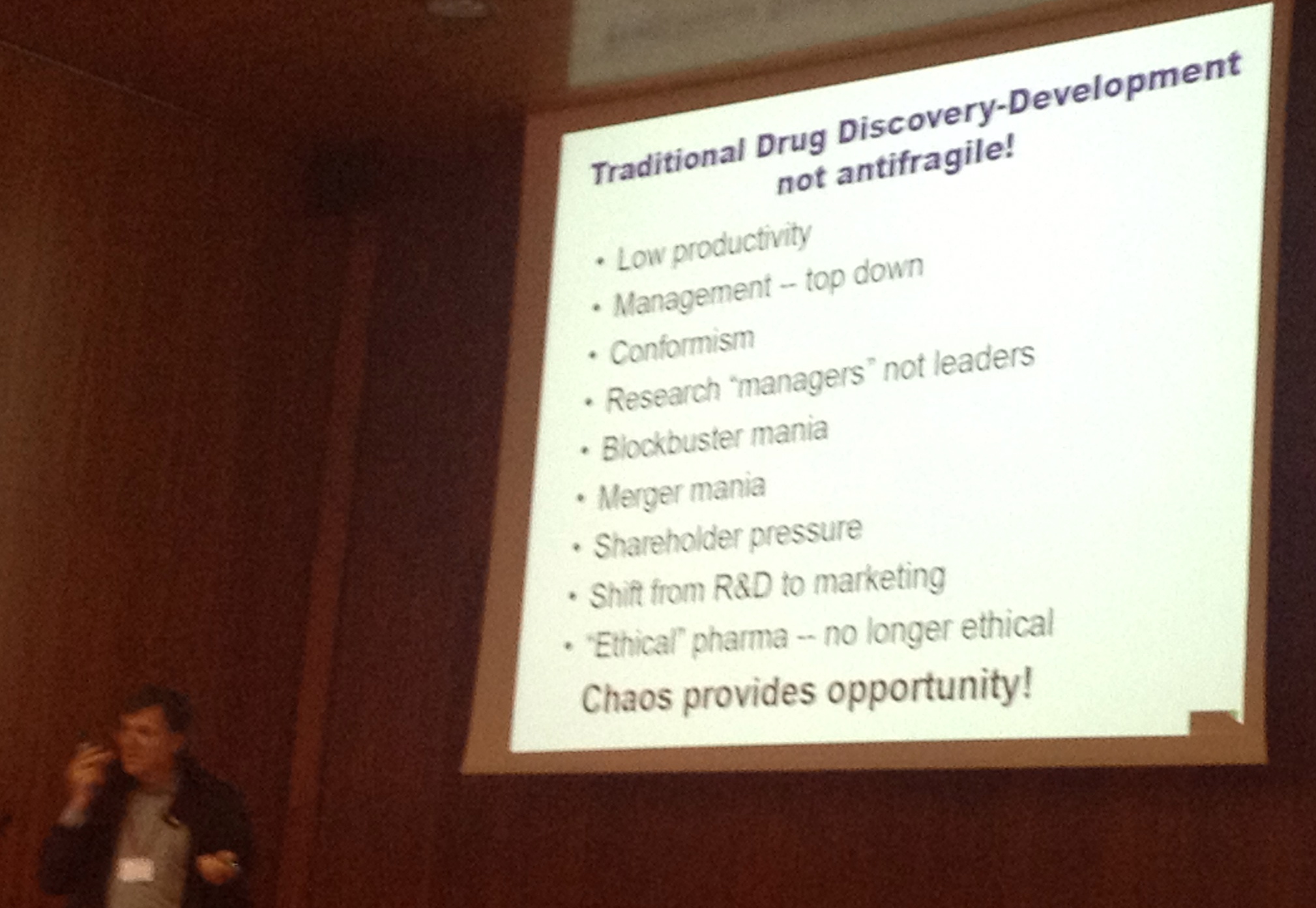
Jim Larrick (Panorama Research, Mountain View, California) gave a typically eclectic, wide-ranging and highly enthusiastic talk on ‘Anti-fragility: Big picture issues in pharmaceutical development’.  He used the “Black Swan” analogy repeatedly to explain how the enterprise funding and pharma research sectors embodied fragile or anti-fragile thinking – with the observation that it was easier to resist black swans (eg: the unexpected) with a raft of small projects, than to have a few big ones.  He also pointed out that the NIH liked big projects – and that a useful alternative name for them was “Not Invented Here”!  Right up there with “Not Real Funding” as the alternative name for our National Research Foundation….

IMG_0133

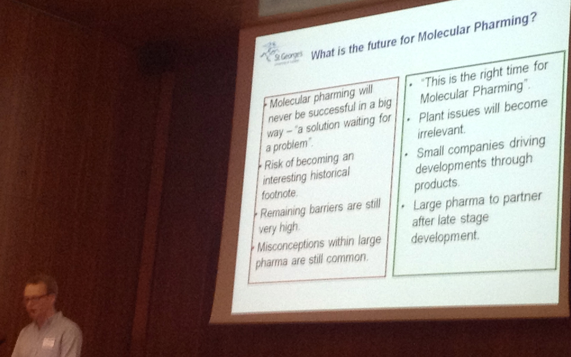
Matthew Paul (St. George’s University of London) presented a set of 15 case studies of commercial paths to introducing molecular farming, which was very interesting to us academic types.  More interesting was the fact that while innovative and protectable technology and products were important to start-ups, the majority of successful ones had their basis in platform development – and the average time from platform to product identification was about five years.  Venture capital firms were considered too greedy for early-stage start-ups, but their involvement later led to stability as their partnering was long term.

Another interesting feature was that many of the successful ventures sold “side products”: for example, Ventria sold cytokines and cosmetic formulations, while KBP sold cell culture reagents.  Several also licenced out technology platforms, but the revenue was not held to be so good.

There were three main indicators of success:

  • Management quality
  • A good lead product
  • Having a panel of products

IMG_0135A good strategy to stay alive was “maximum income / minimum burn” – and he held up the example of Medicago in this regard.  He noted that in the absence of major investment from Big Pharma, Phase 2 trial success was the driver for commercialisation.

PBVAB 5 Verona June 2013: Session 7

3 August, 2013

Suzanne Huddy, a postdoc in our lab, kindly took some notes in a session I moderated at the 5th PBVAB in Verona this year.

Little did she know this is just my way of easing her in to doing this more often…B-)  Thanks, Sue!

Session 7: Manufacturing and Production Systems Developments

Moderator: EP Rybicki

Andreas Schaaf from Greenovation Biotech GmbH presented on “BryotechnologyTM en route to the clinic”, highlighting a production platform based on the moss Physcomitrella patens.  The overriding advantage of this system is that the moss is haploid and therefore genome modification is fairly straight forward with timelines for modifications similar to that of yeast systems.  Physcomitrella patens is also fairly unique since it has a very high occurring rate of homologous recombination (HR).  These traits along with the fact that the genome is sequenced and annotated allow fairly simple customization of the genomic background.  Using this, they have glyco-engineered strains and have removed plantized glycosylation completely.

Other than the products mentioned on their website (www.greenovation.com), they are currently working on α-galactosidase for treating Fabry disease.  Fabry disease is a rare genetic lysosomal storage disorder which results in the accumulation of lipids in the kidney, autonomic nervous system and cardiovascular system cells.  They are also working on the production of recombinant human β glucocerebrosidase for the treatment of Gaucher disease.  Interestingly, these are the same products produced by Protalix Therapeutics.

Stefan Schillberg from the Fraunhofer IME presented on “Co-MoFarm- Contained molecular farming: Controlled contained systems for high yield consistency”.  The CoMoFarm project has been funded for 3.5 years under the European Commission 7th Framework programme.  This project focused on the development of high-yielding plant-based production systems for recombinant proteins.

The presentation initially contrasted the production capability of the various plant platforms employed by this group using both HA (influenza hemagglutinin) and the human M12 antibody as protein products.  The production platforms included Arabidopsis and rice suspension cells, tobacco plants, roots and suspension cells, and moss suspension cultures.  The results presented highlighted the fact that one production platform is not necessarily optimal for all recombinantly expressed proteins, although the traditional tobacco leaves and BY-2 suspension cultures did produce the highest expression levels.  By further optimization of cultivation parameters (including media components), expression levels could be increased by up to 30 fold.  The presentation also showed that expression could also be improved by co-expression of the target protein with a fluorescent marker, DsRed.  In short, this allows the development of higher expressing lines through the non-invasive selection single elite expressing cells by flow-cytometry.  Stephan Schillberg also presented on the groups development of non-invasive monitoring systems for plant cell health and productivity.

The presentation was ended with a comparison on the cost of production of M12 antibody in either tobacco plants or BY-2 cells grown in 200 L bioreactors.  While the cost of producing this product in tobacco plants was less per gram of the product, the time for production in BY-2 cells was much shorter.  Details of the costing can be found at http://comofarm.org/useruploads/files/CoMoFarm_2013-6.pdf, where CoMoFarm have kindly made the presentation given in Verona available.

Pascal Drake from St. George’s University of London presented on “Hydroponic cultivation of tobacco for the production of recombinant pharmaceutical proteins by rhizosecretion”.  This presentation looked at the production and optimization of antibodies and Cyanovirin-N (CV-N) (a cyanobacterial protein which displays virucidal activity) in hydroponically cultivated tobacco plants.  Data was shown that suggested the inclusion of PGRs (plant growth regulators) and a nitrate source in the hydroponic medium could increase the concentration of the protein of interest in the medium.  Hydroponic cultivation has some advantages over traditional cultivation of tobacco plants.  Plants are cultivated in chemically defined media, therefore there is better control over the process and in this way this system approaches cell fermentation processes.  Additionally, fully processed secreted proteins can be harvested over the lifetime of the plant and purification can be simplified since the medium does not contain as many proteins as a whole leaf extract.  A “nifty” way of doing a western blot was also shown- basically, transgenic plants are germinated on nitrocellulose paper; this paper can then be used directly for a western blot since the protein of interest would have been secreted directly from the roots of the plant onto the membrane.  After development of the blot, the presence of the protein is seen in “root-shaped” pattern.

Bertrand Magy from the Institute of Life Sciences at the University catholique de Louvain, Belgium presented on the “Development of suspension cells as a competitive production system for antibodies”.  This research looked at designing an optimized antibody scaffold that can be combined with different variable regions in order to produce high levels of functional antibodies.  Initially, the expression of different IgG isotypes (human, rat and mouse) with the same variable region was investigated in tobacco and Arabidopsis thaliana suspension cells.  Bertrand showed that while antibodies accumulated in the extracellular medium, degradation occurred according to the isotype.  In this case, A. thaliana was also shown to be the better producer.  As is the case with many other cell suspension-based expression, the yield of antibody could be optimized by manipulating the growth medium.  Levels of antibody production of >30 mg/L could be achieved.

Moratorium on using live rinderpest virus lifted for approved research

30 July, 2013

See on Scoop.itVirology News

Benefits of future research should be carefully balanced against potential risks

Paris, 10 July 2013 – A moratorium on using live rinderpest virus for approved research has been lifted by the Food and Agriculture Organization of the United Nations and the World Organisation for Animal Health (OIE).

The moratorium followed the adoption of a Resolution in May 2011 by all OIE Member Countries that urged members to forbid the manipulation of rinderpest virus containing material unless approved by the Veterinary Authority and by FAO and OIE.

The two organizations have now put in place strict criteria and procedures to follow in order to obtain official approval for any research proposals using rinderpest virus and rinderpest virus-containing materials. One of the most crucial requirements is that the research should have significant potential to improve food security by reducing the risk of a reoccurrence of the disease. This procedure replaces an earlier complete ban on handling the virus.

Rinderpest was formally declared eradicated in 2011, but stocks of rinderpest virus continue to exist in laboratories. In June 2012, a moratorium on handling the virus was imposed after an FAO-OIE survey found that the virus continues to be held in more than 40 laboratories worldwide, in some cases under inadequate levels of biosecurity and biosafety.

When rinderpest was officially eradicated, FAO and OIE member countries committed themselves to forbid the manipulation of rinderpest virus-containing material unless approved by the national veterinary authority as well as by FAO and OIE.

Paramyxovirus EM courtesy of Linda Stannard

Thanks to Len Bracher for alerting me to this.

Ed Rybicki‘s insight:

This is an interesting sequel to the eradication of wild rinderpest virus, which I have covered in some detail here on ViroBlogy: see here (https://rybicki.wordpress.com/2010/11/05/rinderpest-gone-but-not-forgotten-yet/) and here (https://rybicki.wordpress.com/2011/08/03/deliberate-extinction-now-for-number-3/).

The article covers an interesting prospect: that it may be possible to use attenuated, safe vaccines against the related peste des petits ruminants virus (PPRV) not only to protect against any resurgence of rinderpest, but also to eradicate this rather nasty virus.

Which is, apparently, spreading at rather an alarming rate, and is an obstacle to small ruminant production (http://www.fao.org/ag/againfo/resources/documents/AH/PPR_flyer.pdf).

So maybe this is “Now for Number 4!” time.

See on www.oie.int

HPV vaccines for South Africa: coming to a school near you!

19 May, 2013

From The Independent Online:

HPV and cervical cancer: courtesy Russell Kightley Media

HPV and cervical cancer: courtesy Russell Kightley Media

“Cape Town – Government will start administering cervical cancer vaccines in schools from February next year, Health Minister Aaron Motsoaledi has announced.

Speaking during the health budget vote debate in the National Assembly on Wednesday, Motsoaledi said government hoped to negotiate lower prices for the vaccine, which treats the Human Papilloma Virus (HPV) – the major cause of cervical cancer among women.

Quoting experts, he said cervical cancer affected 6000 South African women a year, 80 percent of them black. More than half the women affected died of the disease.

While the HPV vaccine presented an opportunity to prevent women from contracting cancer, there were still obstacles to overcome.”

This is a really, really big deal for South Africans – and pity is, the vaccine will not be given to boys, or universally to girls.

Seriously: all the science says that giving it to boys as well limits spread of the viruses far better; not making it universally available will mean all sorts of recriminations around unequal access (read: to less privileged kids ONLY as part of the government programme at first).

But a big step in the right direction!

A Brief History of Influenza

5 April, 2013

See on Scoop.itVirology News

I am TRYING to write an eBook on influenza, which stubbornly refuses to be finished – as part of a sabbatical project, which finished in December 2010.  So, like my History of Virology, I am triall…

Ed Rybicki‘s insight:

I will reprise this post, given a considerable recent spike in interest in it as the new H7N9 Shanghai bird flu starts.  Hopefully to fizzle out, but you never know….

Incidentally, I have an almost-finished iBook (for iPad) on influenza: the first five respondents to this post can trial it for free!

See on rybicki.wordpress.com

Maize streak virus revisited: 25 years on

20 March, 2013
Maize streak virus: photo from 1978

Maize streak virus: photo by Robert G Milne in Cape Town from 1978

Twenty-five years ago, I wrote a brash, naïve little piece entitled “Maize streak virus virus: an African pathogen come home?” for the South African Journal of Science, laying claim to a virus that we had just started working on – Maize streak virus (MSV) – on the basis that it had first been described from this country in 1901, that it was endemic here, and that it still caused major crop losses.  I did this because research on this and related viruses seemed to have moved almost completely offshore, to Europe and the USA, and

“…the most interesting of the viruses that grow all around us have already been whisked away to foreign laboratories; [that] there they have been cloned, sequenced, and had their most intimate details exposed, far from their native shores”. [Yes, I really did write like that back then].

I asked at that time, if we should

“…perhaps be content to supply foreigners with the (pathogenic) fruits of our fields, and to marvel when the answers come filtering back from abroad?”.

I answered myself by saying that

“…prospects for worthwhile research on African geminiviruses, and on any other indigenous pathogens, are at least as good here as anywhere else.  Our facilities are the equal of those abroad, the necessary expertise is certainly not lacking, and the viruses are on our doorstep.”

I’m a little shocked now that I could have said that then: the paper quotes only three pieces of work from our lab, one of them a Masters dissertation and two papers done by my erstwhile supervisors; we had not yet sequenced any virus, let alone a geminivirus, and all we had was brashness and hope.  Indeed, I went on to say the following:

“We are, incidentally, the only research group with access to molecular biological techniques which is actually working on the virus in its natural environment: this is very useful, as with the virus in all its forms and its vector(s) literally on our doorstep, we can rapidly accumulate, identify and characterize distinct isolates for study here or elsewhere.  We hope there will be a little more of the ‘here’, and a little less of the ‘elsewhere’, from now on”.

I outlined what it was that we ambitiously wanted to do – seeing as we had no money, and only one PhD student at the time – as follows:

“…we now have distinctly different genomic maps of three isolates [!] which differ in serology and symptom expression; we have cloned genomic DNA of several more isolates, and can potentially clone and [restriction] map many more.  With this type of work now solidly established, we intend to investigate other biological variants of MSV – and other native cereal geminiviruses – in maize, cereal grains and other members of the Gramineae.  The aim is to explore the genetic diversity of naturally occurring types of MSV and related viruses, and to identify any isolates that appear unusual in terms of symptom expression, serology or transmission.  These would be interesting to map, and potentially useful in recombinational analyses for the fine mapping of determinants of pathogenicity and host range.” [see later]

The article obviously sank without trace: I can find only three citations to it; two of them mine, and the third from a South African maize breeder.  How the overseas labs that I compared us to must have sniggered…actually, I doubt that happened at all; I am sure none of them ever read it!  In retrospect, we really were regarded as a backwater, and as wannabe geminivirologists; I had at least one collaboration request rebuffed with “we don’t feel our work would be advanced by working with you”, and was told “we’re already working on that, so you shouldn’t bother” for a couple of other proposals.

My hubris was not entirely misplaced, however: we did in fact go on to develop into a world-leading MSV and geminivirus molecular virology laboratory; it just took another fifteen years or so!

So where are we, twenty-five years on from my cheeky article?  Much water has flowed under several bridges; I expanded from molecular virology in the 1990s into plant and vaccine biotechnology in the 2000s, while keeping a geminivirus research group going – and we have published and co-published something like 55 peer-reviewed journal articles and several encyclopaedia and book chapters on MSV and other “African streak viruses” alone, let alone another 14 or so articles on other geminiviruses, with some 1200 citations.  We have papers on geminivirus mapping and sequencing, virus diversity, biogeographical variation, quantitation of symptoms, molecular determinants of pathogenicity, recombination, engineering maize for resistance, the use of two of the viruses as gene expression vectors – and cover pictures for Plant Biotechnology Journal and Journal of Virology.

Cover Illustration: J Virol, October 2011, volume 85, issue 20

Cover Illustration: J Virol, October 2011, volume 85, issue 20

I started with one Honours student in 1986, who went on to do a Masters in 1988; we moved on to having one PhD student in the late 1980s to up four PhD students simultaneously in the mid- to late 1990s, and a postdoc at the same time.  The projects went from simple diversity studies of a few viruses using restriction mapping, through the application of PCR, to partial genome sequencing and studying the molecular biology of infectious clones of the viruses, with a very profitable sideline in phylogenetic analyses; we also moved – with Professor Jennifer Thomson – into a parallel track of plant biotechnology, aimed at engineering resistance to MSV in maize.  We added another track early this century, working on similar ssDNA circoviruses of parrots, using all of the expertise we had accumulated on geminiviruses.  We truly work on “circomics” now – the study of small circular genomes – with its subsets “geminiviromics” and “circoviromics”, with a library of literally hundreds of sequenced MSVs and distinct grass mastreviruses and BFDVs.

Geminivirus particle: characteristic doubled icosahedron containing a single ssDNA

Geminivirus particle: courtesy of Russell Kightley Media

The geminiviromics group has pretty much got away from me now; the folk I trained as PhD students in the late 1990s and early 2000s were enthused enough with the field that they have gradually usurped my leadership and supervisory role, and made the field their own.  I still maintain an interest in using Bean yellow dwarf mastrevirus (BeYDV) as an expression vector for “biofarming” purposes; I am also maintaining a project on Beak and feather disease circovirus (BFDV) diversity and plant-made vaccines.  I think we pretty much did what we set out to do – including the brave prediction I made about host range and pathogenicity, which led to some very interesting work on recombination and genome modularity, and the successful engineering of pathogen-derived resistance to MSV.

So I owe some thanks, in retrospect: first, to Barbara von Wechmar, who sparked the interest – and provided isolates, leafhoppers, and expertise.  Second, to Bev Clarke and Fiona Tanzer (aka Hughes), who were brave enough to blaze the trail, and clone our first MSVs – and make one infectious, in the case of Fiona.  Thanks to Wendelin “Popeye” Schnippenkoetter, for your single-minded perseverance in mixing and matching genomes; thanks Kenneth Palmer, for showing the way for transient expression assays in maize cells and engineering MSV as a vector.  Thanks Janet Willment, for mapping replication origins in MSV and expanding us into wheat viruses; thanks Jennifer Thomson for the collaboration, and Fiona and Tichaona Mangwende and Dionne Shepherd for breaking us into maize resistance engineering.  Thanks Christine Rey for the collaboration, and Leigh Berrie for your quiet competence in our detour into South African cassava mosaic virus.  Thanks Darrin (aka Darren) Martin and Eric van der Walt, for so brilliantly exploring MSV diversity, evolution and recombination – and Darrin for endless amusement in the lab, as well as for two completely distinct and invaluable software packages, for symptom quantitation and recombination analysis.  In the present generation, thanks to Suhail Rafudeen and our student Rizwan Syed (and Dionne and Darrin as supernumerary supervisors); thanks Aderito Monjane for doing such a ridiculous amount of work for a superlative PhD; thanks Dionne and Marian, for keeping the maize engineering afloat – and thanks also to Arvind Varsani, for retraining himself from a papillomavaccinologist to a circomicist, and for popping up everywhere.

PLOS Pathogens: Environmental Predictors of Seasonal Influenza Epidemics across Temperate and Tropical Climates

18 March, 2013

See on Scoop.itVirology News

Human influenza infections exhibit a strong seasonal cycle in temperate regions. Recent laboratory and epidemiological evidence suggests that low specific humidity conditions facilitate the airborne survival and transmission of the influenza virus in temperate regions, resulting in annual winter epidemics. However, this relationship is unlikely to account for the epidemiology of influenza in tropical and subtropical regions where epidemics often occur during the rainy season or transmit year-round without a well-defined season. We assessed the role of specific humidity and other local climatic variables on influenza virus seasonality by modeling epidemiological and climatic information from 78 study sites sampled globally. We substantiated that there are two types of environmental conditions associated with seasonal influenza epidemics: “cold-dry” and “humid-rainy”. For sites where monthly average specific humidity or temperature decreases below thresholds of approximately 11–12 g/kg and 18–21°C during the year, influenza activity peaks during the cold-dry season (i.e., winter) when specific humidity and temperature are at minimal levels. For sites where specific humidity and temperature do not decrease below these thresholds, seasonal influenza activity is more likely to peak in months when average precipitation totals are maximal and greater than 150 mm per month. These findings provide a simple climate-based model rooted in empirical data that accounts for the diversity of seasonal influenza patterns observed across temperate, subtropical and tropical climates.

Ed Rybicki‘s insight:

This is really quite a big deal: I blogged recently on the first paper that explored this notion in detail; here we see that paper vindicated, and new data presented.


It is interesting that the virus should have evolved to be spread in this way: in drier cold air in temperate climates, and in warm wet air in more tropical climes.  It also very nicely explains seasonality in influenza transmission.


Now, let’s do something ABOUT it!

See on www.plospathogens.org

14 adults ‘cured’ of killer HIV virus [NOT!!]

16 March, 2013

See on Scoop.itVirology News

TWO weeks after doctors rid a baby of the disease, it appears the treatment has worked on full-grown men and women

Ed Rybicki‘s insight:

You have to hate sub-editors – the people who are tasked, in papers like the Sun, to come up with the most lurid headline possible.


The facts are these: a number of people were treated, soon after infection with HIV-1, with a course of combo ARVs.  For one reason or another, they stopped taking them – and they are, up to seven years out – controlling their virus load to undetectable levels.


Note: they are almost certainly NOT cured; the virus is integrated into their CD4+ T-cells, and is simply quiescent or ticking over at a very low level of expression.


Howevr, it is potentially good news – IF it can be replicated in a wider cohort, and IF people can be caught at an early stage of infection.

See on www.thesun.co.uk

Vaccines: a simple message

28 February, 2013

+MaryMangan over there on Google+ made an interesting point about simple messages to refute the kinds of nonsense promulgated by vaccine denialists, among others.

Here’s my contribution:

Vaccines!

Vaccines!

TMV in mouse lungs: more thoughts and refutations

13 February, 2013

tmv sedimhave been thinking about this paper (see last post), and it and other people’s posts (eg: Tommy Leung’s) have prompted more response.

I note the authors  say the following:

“There is other published literature that challenges the dogma of the strict boundaries between plants and vertebrates for viruses. In non-vertebrate animals, it was shown that plant pathogenic viruses displayed complex interactions with insects, and the transcription and replication of some plant viruses within insects was described [29][32]. In addition, in some cases, insects were found to be affected by plant viruses [33]. Furthermore, it was recently shown that Tomato spotted wilt virus (TSWV) could infect two human cell lines, HeLa and diploid fibroblasts, depending on the expression of a viral polymerase-bound host factor[34]. Additionally, despite plant virus replication was not observed in animals, Cowpea mosaic virus (CPMV), a plant comovirus in the picornavirus superfamily, was able to bind and enter mammalian cells, including endothelial cells, and the binding protein for the virus was identified as a cell-surface form of the intermediate filament vimentin [35]. Furthermore, CPMV was found to persist for several days post oral or intravenous inoculation in a wide panel of body tissues in mice, including in the lung and the liver [36]. Additionally, it was demonstrated that TSWV induced a strong immune response in its insect vector Frankliniella occidentalis [37] and that oral administration of Cowpea severe mosaic virus, Alfalfa mosaic virus and chimeric plant virus particles induced a durable and systemic immune response in mice [38][39]

Yes.  Um. Well.  The “dogma of the strict boundaries between plants and vertebrates for viruses”?  I have been teaching virology for 32 years, and I am not aware of actual DOGMA – as in, “that which has to be believed”.  Rather, there has been the cumulative set of OBSERVATIONS that nothing that anyone has ever isolated out of a plant – and that replicates in it – has infected a vertebrate.  I make that distinction, because there is always the possibility that, as we and others have found with insect viruses, plants can act as a “circulative, non-propagative vector” for insect viruses (for Rhopalosiphum padi aphid virus in barley, from my lab, and Leafhopper A virus in maize) – and if one realises that male mosquitoes, and often also females, feed on plants…you see where I’m going here?  As in, it might well be possible for a virus that multiplies in an insect and also in a vertebrate, to POTENTIALLY be found in a  plant?

In ay case, this is largely beside the point, because the authors get sidetracked into discussing Tomato spotted wilt – which happens to be a plant-adapted bunyavirus, most closely related to insect and vertebrate phleboviruses – “depending on the expression of a viral polymerase-bound host factor”.  Really??  And if it isn’t there?  Does the virus in fact spread?  For that matter, my lab has cell-free translated two aphid picorna-like virus genomes in rabbit reticulocyte lysates, but we made no claim that it could happen in rabbit cells.  Moreover, they make much of the fact that “a plant comovirus in the picornavirus superfamily, was able to bind and enter mammalian cells…[and] was found to persist for several days post oral or intravenous inoculation in a wide panel of body tissues in mice, including in the lung and the liver”.

Yes?  And?  A REALLY stable plant virus was able to bind and enter animal cells, and persist?  The problem with that is…?

We in the virus-like particle vaccine field RELY on the fact that VLPs will be taken up by cells of the immune system in vertebrates, and that they will elicit immune responses – so why is this regarded as a problem?  In fact, TMV has itself been tested as an RNA vaccine delivery system, due to its ability to protect a RNA payload, and get itself delivered into reticulocytes and macrophages – meaning this property has been known for some time, and has not hitherto been seen as a problem!

I think these authors have hyped something that is quite interesting into what THEY regard as a potential problem, for the purposes of getting their article accepted – and I think this needs to be recognised, and that the perceived risks need to be minimised by the knowledgeable.