Posts Tagged ‘electrophoresis’

Gone, but not quite forgotten: the Rybicki teaching pages

6 August, 2015

I have extolled the virtues of the Internet Archive’s Wayback Machine previously, as a magic means of finding material that you probably thought (and sometimes wished) was long lost: in that instance it was my old Ebola news pages.

I now find a new reason to commend its virtues to the skies: I once wrote, on The Guru Cann’s site,

“So how does one even approach the problem of constructing a history of any particular corpus of web-published material?”

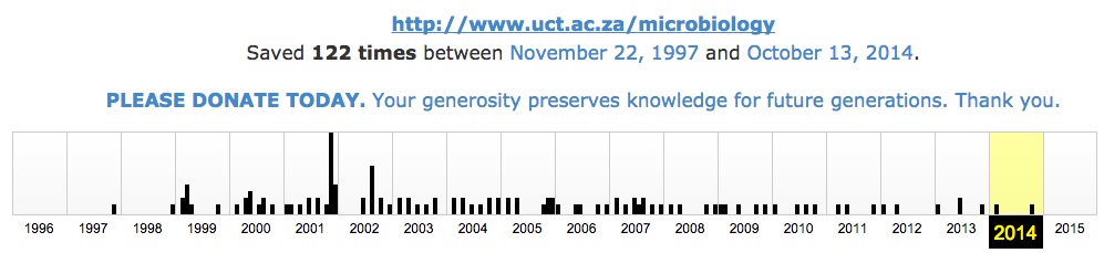
The Wayback Machine, it appears is an answer.  Not THE answer, because there are still holes in its coverage, but here is an example of how many iterations there are of archives of my Web-based PCR Methods teaching pages:

Banners_and_Alerts_and_PCR

Right back to 2004!  The teaching material goes back to 1997, along with my primitive efforts at a Departmental Web page – like the old Department of Microbiology, all my pages are now defunct

Internet_Archive_Wayback_Machine– because our University, in their wisdom, has now decided to switch to Drupal-based web sites, meaning all my old material along with the servers it’s on, is dead.

Defunct.  Deceased.  No longer with us.  Except…

I find, to my joy, that you CAN in fact get to nearly all of it, and backed up as recently as March 2014, via this link:

MCB_-_Online_teaching_material

Might not be completely back from the dead, but it’s a reasonable facsimile – and it means that if anyone was using it, they can continue to do so – while I sort out new versions, and new addresses.

And, of course, finish my book based on it…B-)

Till then!

ELECTROPHORESIS OF THE RABBIT PAPILLOMA VIRUS

21 August, 2012

See on Scoop.itVirology News

This is synchronicity, of a sort, seeing as I recently blogged on whole-virus electrophoresis done in our labs sometime in the late 1970s.  This is a whole lot more complicated, as it happens, but still a useful tool.  If you can get enough virus, that is!

See on www.starhi.com

A bit of viral archeology

20 July, 2012

We were sifting through stuff found in a service room the other day, when I found a box of glass slides – undisturbed since about 1979 or so.  A very interesting box: double- and triple-width microscope slides, coated with dried agarose gel, and stained with Coomassie Brilliant Blue.  With my handwriting on them.  With whole virus electropherograms on them….

Backing up a bit: back in a previous research career, I was a plant virologist who had become an expert, during my MSc project, on physical and serological techniques to do with plant viruses, and the multicomponent isometric bromoviruses in particular.  This included differential, density gradient and analytical centrifugation; methods for purification of virus, capsid protein and genomic nucleic acid (all ssRNA); double-diffusion gel precipitin (=Ouchterlony’s technique) assays; the then new-fangled enzyme-linked immunosorbent assays (ELISA) – and whole-virus agarose gel electrophoresis, and immunoelectrophoresis.

This is mostly published – in my first-ever paper published in 1981, that I was too naive to know I shouldn’t submit to a good journal, so got it into Virology.

However, there were some bits that only ever made it into my Masters write-up – and then only in monochrome.  Here, then, is a little piece of virological and personal history: electropherograms of Brome mosaic bromovirus strains, electrophoresed in 1% agarose on glass slides, then dried down and stained with CBB.  Left all alone, in a drawer, undisturbed from then till now.

It is very easy to see how the three strains on the right have a pI between pH 6.0 and pH 7.5, and that the two on the left and the one on the furthest right seem to be mixtures of differently-charged variants.

Interesting technique, this: it’s a very nice way of characterising and in fact separating virus strains that differ only in a couple of charges in their capsid proteins – for future infectivity assays, if need be, or for preparative purposes by sucrose gradient “zone” electrophoresis, as done here.  The thing about the slide gels, though, is that it is very cheap, very easy, and very quick – ideal for practicals and demonstrations.

I’m going to make my current MSc student characterise his plant-made virus-like particles this way…B-)

Oh, and while we’re archeologising: here is a 30+ year experiment in sedimentation at one gravity, of Tobacco mosaic virus.  Shows why one should stay in one place for a while.  Or not…B-)