Archive for the ‘Evolution’ Category

CCHFV in South Africa

1 February, 2013

I am indebted to the National Institute for Communicable Diseases (NICD) in Johannesburg for their very informative newsletter, from which I culled this.

I would also like to very sincerely congratulate Professor Barry Schoub, a long-time former Director of the NICD, on his  African Society for Laboratory Medicine (ASLM) Lifetime Achievement Award!  Very well deserved.

Crimean-Congo haemorrhagic fever

Two cases of Crimean-Congo haemorrhagic fever (CCHF)  acquired in South Africa have been laboratory confirmed  in January 2013.

On 1 January 2013, a 31-year-old male working as a  game warden on private game ranch near Jagersfontein  (Free State Province) presented with clinical features  suggestive of CCHF. The patient did not report any tick  bites or direct exposure to unprocessed meat or  slaughtering of animals. The Centre for Emerging and  Zoonotic Diseases of the NICD/NHLS confirmed infection  with CCHF virus by PCR and serology testing.

A second case of CCHF was laboratory confirmed on 12  January 2013 in a 44-year-old male hospitalised in  Bloemfontein, Free State Province. He had been on a  farm in Pomfret, North West Province (situated ±5 km  from the border with Botswana), where he was bitten by  a tick. Three days later he developed symptoms, and  presented with fever, rash, conjunctivitis and pharyngitis.  No laboratory-confirmed cases were identified in 2011- 2012.

Human CCHF cases have been reported annually  from South Africa since 1981, when it was first  recognised in the country; between 0 and 20 cases of  CCHF are diagnosed each year. Through nearly thirty  years of passive surveillance, a total of 187 cases has  been laboratory confirmed. Although cases have been  reported from all of the nine provinces, more than half of  the cases originate from the semi-arid areas of Northern  Cape Province (31.5% of cases) and Free State Province  (23% of cases).

CCHF infection is generally asymptomatic in many species  of wildlife (including antelope) and livestock animals  (including cattle, sheep, goats, hares and ostriches).  Humans  become  infected  sporadically  by  ticks,  particularly  Hyalomma ticks, which are both reservoirs  and vectors for CCHF virus. Other modes of transmission  include direct contact with blood/tissues of infected  animals, and in the case of healthcare workers, through  direct contact with the blood/tissue of infected patients;  nosocomial outbreaks are well described and have been  associated with high mortality rates. Disease may be  severe in people, with case-fatality rates reported as 3 –  30% across various studies.

Detailed information for healthcare workers regarding  CCHF can be found on the NICD website  http:// http://www.nicd.ac.za/ (see General Public FAQ, or Health Workers FAQs here).

And so it went – 2012, that is

14 December, 2012

…like a rocket…flashed past; I’m still emotionally in August or so!

I meant to do some more substantive posts instead of only copying Scoop.it Virology News posts here; however, the best-laid plans and such, and I didn’t.  I will in 2013, though – and there will be an iBook coming or possibly even two (influenza and PCR), so I will use this forum to announce glad tidings.

Then there’s the ZA Virus [=Zombie Apocalypse, obviously] novel, and Green Vaccines, and…OK, getting ahead of myself here!

Thanks for the support and readership, I hope everyone has a good solstice break!

Best,

Ed

PS: some access stats for 2012 for you.  Looks like the only places that DON’T access ViroBlogy are parts of central and west Africa, central Asia and Greenland.

Screenshot 14:12:2012 12:11

Screenshot 14:12:2012 12:09


Screenshot 14:12:2012 12:20

Together, we can do more….

17 October, 2012

It gives me great delight to pass on some news about an old friend: I have co-authored two papers with the Pappus (husband and wife), and have maintained a long association with Hanu as a favoured referee for Archives of Virology; he has gone on to achieve some distinction at Washington State University – and recently to have made a fundamental discovery in plant virology.  I thank Eric Sorenson of the Washington State Magazine for sending me this.

Viral alliances overcome plant defenses, according to newly published WSU research

Contact:
Hanu Pappu, professor and chair of plant pathology, Washington State University, 509-335-3752, hrp@wsu.edu

PULLMAN, Wash. – Washington State University researchers have found that viruses will join forces to overcome a plant’s defenses and cause more severe infections.

“These findings have important implications in our ability to control these viruses,” says Hanu Pappu, Sam Smith Distinguished Professor of Plant Virology and chair of WSU’s Department of Plant Pathology. “Mixed infections are quite common in the field, and now we know that viruses in these mixed infections are helping each other at the genetic level to overcome host defenses and possibly lead to the generation of new viruses.”

Pappu publishes his findings in the latest issue of the journal PLOS ONE. Joining him are Ph.D. student Sudeep Bag and Neena Mitter, associate professor at Australia’s University of Queensland.

The researchers focused on iris yellow spot virus and tomato spotted wilt virus after Bag discovered that, when they infect the same plant, they helped each other overcome a plant’s defense response. With Mitter’s help and sophisticated molecular techniques, Bag found both viruses dramatically changed their genetic expression, breaking down the plant’s defenses and leading to more severe disease.

Bag also found that genes from the tomato spotted wilt virus seemed to “aid and abet” iris yellow spot virus as it spread throughout the plant and caused more disease.

Growers should take this phenomenon into account, says Pappu, with broader management tactics that target more than one virus and possible variations.

The research was funded in part by the Specialty Crops Research Initiative of the National Institute of Food and Agriculture, a branch of the U.S. Department of Agriculture.

The paper, “Complementation between Two Tospoviruses Facilitates the Systemic Movement of a Plant Virus Silencing Suppressor in an Otherwise Restrictive Host,” can be found athttp://dx.plos.org/10.1371/journal.pone.0044803.

PS: the Pappus cook REALLY good food – as I discovered in Florida, at Chuck Niblett’s house, back in 1996 or so….

White death: A diabolical pact between an insect and two viruses

22 August, 2012

See on Scoop.itVirology News

This is actually an article in The Economist from 2007 – forwarded to me by a Professor of Philosophy, as it happens, and which has mouldered on my desk lo, these past five years.  Thanks David Benatar!

“Whiteflies are pests in every continent that they are found in—and they are found in every continent except Antarctica. They cause damage directly, by consuming plant juices, and indirectly, by spreading viral diseases. But Liu Shusheng, of Zhejiang University, in Hangzhou, and his colleagues have found a strain of the species that delivers a double whammy. Not only does it spread diseases, but it is also vastly more successful when it lives on plants infected with the diseases in question [tomato yellow leafcurl and tobacco curly shoot begomoviruses, both ssDNA geminiviruses]  than when it subsists on healthy plants.”

This is a fascinating example of just why it is that certain vector-virus-host combinations can lead to success of the vector, and increased spread of the virus.  Basically,

“…type B insects lived six times longer on infected plants than uninfected ones, and their population per infected plant might rise as high as 13 times that on an uninfected one”

This means that geminivirus infection of host plants actually gives a survival advantage to the insects which transmit them.  Simple if unfortunate!!

See on www.economist.com

Papillomavirus and HIV: a nasty combination

17 August, 2012

I started working on human papillomaviruses (HPVs) some 22 years ago, back at the dawn of PCR: I helped my then-new major collaborator (and wife of 2 years), Anna-Lise Williamson, design some degenerate primers for amplifying as wide a range as possible of high-risk HPVs from cervical biopsy samples.  These worked pretty well, and are still highly useful for the purpose, despite the many novel types found since then.

We went on to do another two papers together on looking at variation and typing of HPVs via PCR and and sequencing, then took a deviation into making candidate vaccines for HPV and HIV.  Anna-Lise carried on with surveilling for HPVs, however, and has ended up with a WHO Regional Laboratory for HPV work.  She also started working on HPV infections in HIV-infected women: work on a study cohort showed that while HIV-free women usually had only 1 HPV type, the 109 HIV-infected often were infected with multiple HPV types.  In association with Anna Salimo in my lab, we started a deep sequencing pilot study on the sample with the most HPVs.  This turned this into a regional study, with help on assembling and interpreting sequence data from Prof Johan Burger’s lab at the University of Stellenbosch, and it was revelatory: while a commercial kit could detect 12 HPV types in one sample, next-gen sequencing found 16.

We went on to do PCR on all 109 samples in the cohort with specific primers for the types not found by the kit, and showed prevalences up to 15% in the HIV-infected group.  This is an important result, because otherwise-innocuous HPV types that do not show up in normal women, may well be associated with disease in the HIV-infected – and will probably not be protected against by the current HPV vaccines.

We continue to do work on these samples, and it will be very interesting to see what the new methodologies show up.  Especially as sequencing becomes cheaper, and we can do more samples…!  Meantime, we have published the pilot study:

Next-generation sequencing of cervical DNA detects human papillomavirus types not detected by commercial kits

Tracy L MeiringAnna T SalimoBeatrix CoetzeeHans J MareeJennifer MoodleyInga I HitzerothMichael-John FreeboroughEd P Rybicki and Anna-Lise Williamson

Virology Journal 2012, 9:164 doi:10.1186/1743-422X-9-164

Published: 16 August 2012

Abstract (provisional)

Background

Human papillomavirus (HPV) is the aetiological agent for cervical cancer and genital warts. Concurrent HPV and HIV infection in the South African population is high. HIV positive (+) women are often infected with multiple, rare and undetermined HPV types. Data on HPV incidence and genotype distribution are based on commercial HPV detection kits, but these kits may not detect all HPV types in HIV + women. The objectives of this study were to (i) identify the HPV types not detected by commercial genotyping kits present in a cervical specimen from an HIV positive South African woman using next generation sequencing, and (ii) determine if these types were prevalent in a cohort of HIV-infected South African women.

Methods

Total DNA was isolated from 109 cervical specimens from South African HIV + women. A specimen within this cohort representing a complex multiple HPV infection, with 12 HPV genotypes detected by the Roche Linear Array HPV genotyping (LA) kit, was selected for next generation sequencing analysis. All HPV types present in this cervical specimen were identified by Illumina sequencing of the extracted DNA following rolling circle amplification. The prevalence of the HPV types identified by sequencing, but not included in the Roche LA, was then determined in the 109 HIV positive South African women by type-specific PCR.

Results

Illumina sequencing identified a total of 16 HPV genotypes in the selected specimen, with four genotypes (HPV-30, 74, 86 and 90) not included in the commercial kit. The prevalence’s of HPV-30, 74, 86 and 90 in 109 HIV positive South African women were found to be 14.6 %, 12.8 %, 4.6 % and 8.3 % respectively.

Conclusions

Our results indicate that there are HPV types, with substantial prevalence, in HIV positive women not being detected in molecular epidemiology studies using commercial kits. The significance of these types in relation to cervical disease remains to be investigated.

I thank Russell Kightley Media for use of the HPV and cervical cancer graphic.

Hidden evolutionary complexity of Nucleo-Cytoplasmic Large DNA viruses of eukaryotes

15 August, 2012

See on Scoop.itVirology and Bioinformatics from Virology.ca

The Nucleo-Cytoplasmic Large DNA Viruses (NCLDV) constitute an apparently monophyletic group that consists of at least 6 families of viruses infecting a broad variety of eukaryotic hosts. A comprehensive genome comparison and maximum-likelihood reconstruction of the NCLDV evolution revealed a set of approximately 50 conserved, core genes that could be mapped to the genome of the common ancestor of this class of eukaryotic viruses.

Results
We performed a detailed phylogenetic analysis of these core NCLDV genes and applied the constrained tree approach to show that the majority of the core genes are unlikely to be monophyletic. Several of the core genes have been independently acquired from different sources by different NCLDV lineages whereas for the majority of these genes displacement by homologs from cellular organisms in one or more groups of the NCLDV was demonstrated.

Conclusions
A detailed study of the evolution of the genomic core of the NCLDV reveals substantial complexity and diversity of evolutionary scenarios that was largely unsuspected previously. The phylogenetic coherence between the core genes is sufficient to validate the hypothesis on the evolution of all NCLDV from a common ancestral virus although the set of ancestral genes might be smaller than previously inferred from patterns of gene presence-absence.


Interesting stuff!  Strengthens my contention that  “…a virus is an infectious acellular entity composed of compatible genomic components derived from a pool of genetic elements” – https://rybicki.wordpress.com/2012/07/10/a-feeling-for-the-molechism-revisited/

Baculovirus image from my collection

See on www.virologyj.com

Adaptive Changes in Alphavirus mRNA Translation Allowed Colonization of Vertebrate Hosts

10 August, 2012

See on Scoop.itVirology and Bioinformatics from Virology.ca

“Genetic, phylogenetic, and biochemical data presented here support an evolutionary scenario for the natural history of alphaviruses, in which the acquisition of DLP structure in their mRNAs probably allowed the colonization of vertebrate host and the consequent geographic expansion of some of these viruses worldwide.”


I have taught for some time now that the evolution of many mammalian viruses must have involved adaptation of originally (and sometimes still) insect-infecting agents – given that insects crawled out onto dry land quite a long time before vertebrates did.  This is a nice illustration of that.  Pity I don’t teach anymore B-(

See on jvi.asm.org

A feeling for the Molechism* – revisited

10 July, 2012

This is an update of a post I did on Alan Cann’s MicrobiologyBytes back in 2007, before i started ViroBlogy: I am doing this because (a) it’s mine, (b) I want to update it – and the MB version is archived, so I can’t.  So here we are again:

I think it’s permissible, after working on your favourite virus for over 20 years, to develop some sort of feeling for it: you know, the kind of insight that isn’t directly backed up by experiment, but that may very well be right. Or not – but in either case, it would take a deal of time and a fair bit of cash to prove or disprove, and would have sparked some useful discussion in the meantime. And then, of course, the insights you have into (insert favourite virus name here) – if correct – can usually be extended into the more general case, and if you are sufficiently distinguished, people may actually take them on board, and you will have contributed to Accepted Wisdom.

I can’t pretend – at least, outside of my office – to any such Barbara McClintock-like distinction; however, I have done a fair bit of musing on my little sphere of interest as it relates (or not) to the State of the Viral Universe, and I will share some of these rambles now with whomever is interested.

I have been in the same office now, and teaching the same course, more or less, for 32-odd years. In that time I have worked on the serology and epidemiology of the bromoviruses, cucumovirus detection, potyvirus phylogeny, geminivirus diversity and molecular biology, HIV and papillomavirus genetic diversity, and expressing various bits of viruses and other proteins in plants and in insect cells. However, much of my interest (if not my effort) in that time has been directed towards understanding how grass-infecting mastreviruses in particular interact with their environment and with each other, in the course of their natural transmission cycle.

Maize streak virus

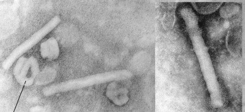
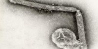
Maxwell’s Demon (left, lower) and Martian Face (right, upper) visible on a MSV virion

Fascinating little things, mastreviruses: unique geminate capsid architecture, and at around a maximum of 2.8 kb of single-strand circular DNA, we thought they were the smallest DNA genomes known until the circoviruses and then the zoo of anello- and anello-like viruses were discovered. Their genomes code for only 4 proteins – two replication-associated, one movement and one capsid – yet we have managed to work on just one subgroup of mastrevirus species for 27 years, without exhausting its interest – at least, to us… (see PubMed list here). We also showed that one could see Martian faces quite distinctly on virions – and possibly even Maxwell’s Demon. But I digress….

Maize streak

Severe symptoms of MSV on sweetcorn

We have concentrated on the “African streak viruses” – related species Maize streak virus, Panicum streak virus, Digitaria streak virus, Sugarcane streak virus and friends – for two very simple reasons:
1. They occur in Africa, near us, and nowhere else;
2. Maize streak virus is the worst viral pathogen affecting maize in Africa.

So we get situational or niche advantage, and we get to work on an economically-important pathogen. One that was described – albeit as “…not of…contagious nature” – as early as 1901, no less.

Maize streak virus

Maize streak virus or MSV, like its relatives, is obligately transmitted by a leafhopper (generally Cicadulina mbila Naudé): this means we have a three-party interaction – of virus-host-vector – to consider when trying to understand the dynamics of its transmission. Actually, it’s more complicated than that: we have also increasingly to consider the human angle, given that the virus disease affects mainly the subsistence farming community in Africa, and that human activity has a large influence on the spread of the disease. So while considering just the virus – as complicated as that is – we have to remember that it is only part of the whole picture.

So how complicated is the virus? At first sight, not very: all isolates made from severe maize infections share around 97% of their genome sequence. However, however…that 3% of sequence variation hides a multitude of biological differences, and there is a range of relatives infecting grasses of all kinds, some of which differ by up to 35% in genome sequence. Moreover, maize is a crop plant first introduced to Africa a maximum of 500 years ago, so it is hardly a “natural” host – yet, all over Africa, it is infected by only a very narrow range of virus genotypes, from a background of very wide sequence diversity available.

So here’s an insight:

the host selects the virus that replicates best in it.

And lo, we found that in the Vaalharts irrigation area in the north of South Africa that the dominant virus genotype in winter wheat was a different strain – >10% sequence difference – to the one in the same field, in summer maize. Different grass species also have quite different strains or even species of streak viruses best adapted to them.

DendrogramNot all that profound a set of observations, perhaps, but they lead on to another insight:

streak viruses travel around as a cloud of variants or virus complex.

Not intuitively obvious, perhaps…but testable, and when we did, we found we were right: cloning virus genomes back out of maize or from a grass infected via leafhoppers gave a single predominant genotype in each case, with a number of other variants present as well. Looking further, we discovered that even quite different viruses could in fact trans-replicate each other: that is, the Rep/RepA complex of one virus could facilitate the replication of the genome of a virus differing by up to 35% in DNA sequence. We have also – we think – made nonsense of the old fancy that you could observe “host adaptation” of field isolates of MSV: we believe this was due to repeated selection by a single host genotype from the “cloud” of viruses transmitted during the natural infection cycle.

So, insight number three:

there is a survival benefit for the viruses in this strategy.

This is simple to understand, really, and relates to leafhopper biology as well as to host: the insects move around a lot, chasing juicy grasses, and it would be an obvious advantage to the streak virus complex to be able to replicate as a complex in each different host type – given that different virus genotypes have differential replication potential in the various backgrounds. This is quite significantly different, incidentally, to what happens with the very distantly-related (in terms of geological time) begomoviruses, or whitefly-transmitted geminiviruses: these typically do not trans-replicate each other across a gap of more than 10% of sequence difference.

Boring, I hear you say, but wait…. Add another factoid in, and profound insights start to emerge. In recent years, the cloud of protégés or virologist complex around me has accumulated to critical mass, and one of the most important things to emerge – apart from some frighteningly effective software for assessing recombination in viral genomes, which I wish he’d charge for – was Darren Martin’s finding that genome recombination is rife among African streak viruses. This was unexpected, given the expectation that DNA viruses simply don’t do that sort of thing; that promiscuous reassortment of components between genomes is a hallmark of RNA viruses. Makes sense in retrospect (an exact science), however, because of the constraints on DNA genomes: how else to explore sequence space, if the proof-reading is too good? And if you travel in a complex anyway…why not swap bits for biological advantage?

MSV web

Linkage map of the MSV genome, showing what interacts with what

So Darren swapped a whole lot of bits, in a tour-de-force of molecular virology, to create some 54 infectious chimaeric MSV genomes – and determined that

The pathogenicity of chimeras was strongly influenced by the relatedness of their parental viruses and evidence was found of nucleotide sequence-dependent interactions between both coding and intergenic regions“.

In other words –new insight:

the whole genome is a pathogenicity determinant, and bits of it interact with other bits in unexpected ways.

At this point you could say “Hey, all his insights are in fact hypotheses!” – and you would be partially correct, except for

Profound Insight No. 1hypotheses are the refuge of the linear-thinking.

Or its variant, found on my office wall:

“**c* the hypotheses, let’s just discover something”. I also have

“If at first you don’t succeed, destroy all evidence that you tried” and a number of exotic beer bottle labels on my wall – but I digress….

As an aside here, I am quite serious in disliking hypothesis-driven science: I think it is a irredeemably reductionist approach, which does not easily allow for Big Picture overviews, and which closes out many promising avenues of investigation or even of thought. And I teach people how to formulate them so they can get grants and publications in later life, but I still think HDS is a tyranny that should be actively subverted wherever possible.

Be all this as it may, now follows

Profound Insight No. 2genome components may still be individually mobile even when covalently linked.

Now take a moment to think on this: recombination allows genes to swap around inside genetic backgrounds so as to constitute novel entities – and the “evolutionary value of exchanging a genome fragment is constrained by the number of ways in which the fragment interacts with the rest of the genome*“. Whether or not the genome is RNA, DNA, in one piece or divided. All of a sudden, the concept of a “virus genome” as a gene pool rather than a unitary thing becomes obvious – and so does the reductionism inherent in saying “this single DNA/RNA sequence is a virus”.

So try this on for size for a brand-new working definition of a virus – and

Profound Insight No. 3a virus is an infectious acellular entity composed of compatible genomic components derived from a pool of genetic elements.

Sufficiently paradigm-shifting for you? Compare it to more classical definitions – yes, including one by AJ Cann, Esq. – and see how much simpler it is. It also opens up the possibility that ANY virus as currently recognised is simply an operational assembly of components, and not necessarily the final article at all.

Again, my favourite organisms supply good object examples: the begomoviruses – whitefly-transmitted geminiviruses –

  • may have one- or two-component genomes;
  • some of the singleton A-type components may pick up a B-type in certain circumstances;
  • some doubletons may lose their B without apparent effect in model hosts;
  • some A components may apparently share B components in natural infections;
  • the A and B components recombine like rabbits with cognate molecules (or Bs can pick up the intergenic region from As);
  • in many cases have one or more satellite ssDNAs (β DNA, or nanovirus-related components) associated with disease causation;

…and so on, and on…. An important thing to note here is the lab-rat viruses – those isolated early on, and kept in model plant species in greenhouses – often don’t exhibit any of these strangenesses, whereas field-isolated viruses often do.

Which tells you quite a lot about model systems, doesn’t it?

But this is not only true of plant viruses: the zoo of ssDNA anello-like viruses found in humans and in animals – with several very distantly-related viruses to be found in any individual, and up to 80% of humans infected – just keeps on getting bigger and weirder. Added to the original TT virus – named originally for the initials of the Japanese patient from whom it was isolated, and in a post hoc exercise of convoluted logic, named Torque teno virus (TTV) [why don’t people who work with human or animal viruses obey ICTV rules??] – are now Torque teno minivirus (TTMV) and “small anellovirus” SAV) – all of which have generic status. And all of which may be the same thing – as in, TTVs at a genome size of 3.6–3.8 kb may give rise to TTMVs (2.8-29 kb) and SAVs (2.4-2.6 kb) as deletion mutants as part of a population cloud, where the smaller variants are trans-replicated by the larger. Thus, a whole lot of what are being described as viruses – without fulfilling Koch’s Postulates, I might point out – are probably only “hopeful monsters” existing only as part of a population. Funnily enough, this sort of thing is much better explored in the ssDNA plant virus community, given that working with plant hosts is so much easier than with human or animal.

And now we can go really wide, and attempt to be profound on a global scale: it should not have escaped your notice that the greatest degree of diversity among organisms on this planet is that of viruses, and viruses that are found in seawater in particular. There is a truly mind-boggling number of different viruses in just one ml of seawater taken from anywhere on Earth, which leads respectable authors such as Curtis Suttle to speculate that viruses almost certainly have a significant influence on not only populations of all other marine organisms, but even on the carbon balance of the world’s oceans – and therefore of the planet itself.

Which leads to the final, and most obvious,

Profound Insight (No. 4)in order to understand viruses, we should all be working on seawater…. 

That is where the diversity is, after all; that is where the gene pool that gave rise to all viruses came from originally – and who knows what else is being

Hypolith – cyanobacteria-derived, probably – under a piece of Namib quartzite from near Gobabeb Research Station

cooked up down there?

And this is the major update: not only have I managed to get funded for a project on “Marine Viromics” from our local National Research Foundation – a process akin to winning the lottery, and about as likely to succeed – I am also collaborating with friends and colleagues from the Institute for Microbial Biotechnology and Metagenomics at the University of the Western Cape on viruses in desert soils, and associated with hypoliths– or algal growths found under quartzite rocks in extreme environments.

Thus, I shall soon be frantically learning how to deal with colossal amounts of sequence data, and worse, learning how to make sense of it.  We should have fun!

——————————————————————————————————————–

* And as a final curiosity, I find that while I – in common with the World Book Encyclop[a]edia and Learning Resources – take:mol|e|chism or mol|e|cism «MOL uh KIHZ uhm», noun. to mean any virus, viewed as an infective agent possessing the characteristics of both a living microorganism and a nonliving molecule; organule.
[molechism < mole(cule) + ch(emical) + (organ)ism; molecism < molec(ule) + (organ)ism] –
There is another meaning… something to do with sacrifice of children and burning in hellfire eternally. This is just to reassure you that this is not that.

How Seanan McGuire Perfected Her Fictional Zombie Virus

27 June, 2012

See on Scoop.itVirology News

When Seanan McGuire set out to write her Newsflesh trilogy, she went to some extreme lengths to make her zombie virus as realistic as possible….


OK, ONE more on zombie apocalyspes (thanks @gussilber)!  Except she didn’t: a quote-

“when those two viruses met, they had babies, and what you got was a shifting-antigen flu that does not leave the body under any circumstances but is capable of turning into something that converts human tissue into more of the virus. And that’s how we got Kellis-Amberlee, which makes zombies” – don’t make it for me.  Human tissue turning into viruses = bloody slime, NOT walking corpses!  Rabies: now THERE’S yer zombie virus!

See on www.wired.com

Five Mutations Make H5N1 Airborne | The Scientist

23 June, 2012

See on Scoop.itVirology News

“After more than 6 months of heated discussion, the second group that succeeded in making the H5N1 avian flu transmissible between ferrets, considered a good model for human transmission, has published its results. The paper, which came out today (June 21) in Science, demonstrates that only five mutations are needed to confer this aerosol transmissibility among mammals, and that re-assortment between different types of viruses—a technique used by the other group, which published its results last month in Nature—is not necessary.

Said Fouchier in a press conference “We both find … loss of glycosylation at the tip of the HA molecule, and this loss of glycosylation seems to increase the receptor binding specificity of the HA”. And though not all the mutations identified in the two studies match, “the mutations that are not identical still have a similar phenotypic trait,” he added.”


So this is what all the fuss was about?  This is what the NSABB did not want everyone to know?  How could they POSSIBLY think that the international virology and infectious disease community should be kept in the dark about this?  What this work has done has pointed the way along a path that will lead us to understand why and how influenza viruses change in order to more effectively get transmitted when they switch hosts – which is a good thing, surely.

And yet all they see is bioterrorism.

See on the-scientist.com